14
views
0
recommends
+1 Recommend
0 collections
    0
    shares
      • Record: found
      • Abstract: found
      • Article: found
      Is Open Access

      Influence of BOLD Contributions to Diffusion fMRI Activation of the Visual Cortex

      research-article

      Read this article at

      Bookmark
          There is no author summary for this article yet. Authors can add summaries to their articles on ScienceOpen to make them more accessible to a non-specialist audience.

          Abstract

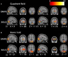
          Reliance on the hemodynamic response as a surrogate marker of neural activity imposes an intrinsic limit on the spatial specificity of functional MRI. An alternative approach based on diffusion-weighted functional MRI (DfMRI) has been reported as a contrast less reliant on hemodynamic effects, however current evidence suggests that both hemodynamic and unique neural sources contribute to the diffusion signal. Here we compare activation patterns obtained with the standard blood oxygenation level-dependent (BOLD) contrast to DfMRI in order to gain a deeper understanding of how the BOLD proportion contributes to the observable diffusion signal. Both individual and group-level activation patterns obtained with DfMRI and BOLD to a visual field stimulation paradigm were analyzed. At the individual level, the DfMRI contrast showed a strong, positive relationship between the volumes of cortex activated in response to quadrant- and hemi-field visual stimulation. This was not observed in the corresponding BOLD experiment. Overall, the DfMRI response indicated less between-subject variability, with random effects analyses demonstrating higher statistical values at the peak voxel for DfMRI. Furthermore, the spatial extent of the activation was more restricted to the primary visual region for DfMRI than BOLD. However, the diffusion signal was sensitive to the hemodynamic response in a manner dependent on experimental manipulation. It was also limited by its low signal-to-noise ratio (SNR), demonstrating lower sensitivity than BOLD. Together these findings both support DfMRI as a contrast that bears a closer spatial relationship to the underlying neural activity than BOLD, and raise important caveats regarding its utilization. Models explaining the DfMRI signal change need to consider the dynamic vascular contributions that may vary with neural activity.

          Related collections

          Most cited references43

          • Record: found
          • Abstract: found
          • Article: not found

          A unified statistical approach for determining significant signals in images of cerebral activation.

          We present a unified statistical theory for assessing the significance of apparent signal observed in noisy difference images. The results are usable in a wide range of applications, including fMRI, but are discussed with particular reference to PET images which represent changes in cerebral blood flow elicited by a specific cognitive or sensorimotor task. Our main result is an estimate of the P-value for local maxima of Gaussian, t, chi(2) and F fields over search regions of any shape or size in any number of dimensions. This unifies the P-values for large search areas in 2-D (Friston et al. [1991]: J Cereb Blood Flow Metab 11:690-699) large search regions in 3-D (Worsley et al. [1992]: J Cereb Blood Flow Metab 12:900-918) and the usual uncorrected P-value at a single pixel or voxel. Copyright (c) 1996 Wiley-Liss, Inc.
            Bookmark
            • Record: found
            • Abstract: not found
            • Article: not found

            Spatial registration and normalization of images

              Bookmark
              • Record: found
              • Abstract: found
              • Article: not found

              What does fMRI tell us about neuronal activity?

              In recent years, cognitive neuroscientists have taken great advantage of functional magnetic resonance imaging (fMRI) as a non-invasive method of measuring neuronal activity in the human brain. But what exactly does fMRI tell us? We know that its signals arise from changes in local haemodynamics that, in turn, result from alterations in neuronal activity, but exactly how neuronal activity, haemodynamics and fMRI signals are related is unclear. It has been assumed that the fMRI signal is proportional to the local average neuronal activity, but many factors can influence the relationship between the two. A clearer understanding of how neuronal activity influences the fMRI signal is needed if we are correctly to interpret functional imaging data.
                Bookmark

                Author and article information

                Contributors
                Journal
                Front Neurosci
                Front Neurosci
                Front. Neurosci.
                Frontiers in Neuroscience
                Frontiers Media S.A.
                1662-4548
                1662-453X
                28 June 2016
                2016
                : 10
                : 279
                Affiliations
                [1] 1Hotchkiss Brain Institute and Department of Radiology, University of Calgary Calgary, AB, Canada
                [2] 2Centre for Advanced Imaging, The University of Queensland St. Lucia, QLD, Australia
                [3] 3Queensland Brain Institute, The University of Queensland St. Lucia, QLD, Australia
                [4] 4Centre for Clinical Research, The University of Queensland Brisbane, QLD, Australia
                [5] 5School of Psychology and Counselling, Faculty of Health, Queensland University of Technology Kelvin Grove, QLD, Australia
                Author notes

                Edited by: Yogesh Rathi, Harvard Medical School, USA

                Reviewed by: Jean-Baptiste Poline, University of California Berkeley, USA; Kevin C. Chan, University of Pittsburgh, USA; Lipeng Ning, Brigham and Women's Hospital, USA

                *Correspondence: Rebecca J. Williams r.j.williams@ 123456ucalgary.ca

                This article was submitted to Brain Imaging Methods, a section of the journal Frontiers in Neuroscience

                †These authors have contributed equally to this work.

                Article
                10.3389/fnins.2016.00279
                4923189
                27445654
                6f95f7ef-e6bd-4d65-b83e-b626f94aad66
                Copyright © 2016 Williams, Reutens and Hocking.

                This is an open-access article distributed under the terms of the Creative Commons Attribution License (CC BY). The use, distribution or reproduction in other forums is permitted, provided the original author(s) or licensor are credited and that the original publication in this journal is cited, in accordance with accepted academic practice. No use, distribution or reproduction is permitted which does not comply with these terms.

                History
                : 07 March 2016
                : 06 June 2016
                Page count
                Figures: 8, Tables: 2, Equations: 1, References: 62, Pages: 15, Words: 11537
                Categories
                Neuroscience
                Original Research

                Neurosciences
                functional mri,fmri,bold,diffusion mri,dfmri,functional imaging,fmri contrast,visual cortex
                Neurosciences
                functional mri, fmri, bold, diffusion mri, dfmri, functional imaging, fmri contrast, visual cortex

                Comments

                Comment on this article