142
views
0
recommends
+1 Recommend
0 collections
    0
    shares
      • Record: found
      • Abstract: found
      • Article: found
      Is Open Access

      Polyglycerolsulfate Functionalized Gold Nanorods as Optoacoustic Signal Nanoamplifiers for In Vivo Bioimaging of Rheumatoid Arthritis

      research-article

      Read this article at

      Bookmark
          There is no author summary for this article yet. Authors can add summaries to their articles on ScienceOpen to make them more accessible to a non-specialist audience.

          Abstract

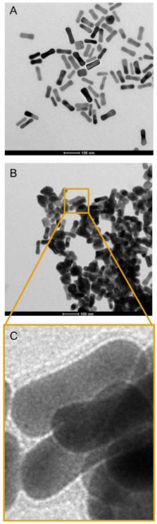
          We have synthesized a targeted imaging agent for rheumatoid arthritis based on polysulfated gold nanorods. The CTAB layer on gold nanorods was first replaced with PEG-thiol and then with dendritic polyglycerolsulfate at elevated temperature, which resulted in significantly reduced cytotoxicity compared to polyanionic gold nanorods functionalized by non-covalent approaches. In addition to classical characterization methods, we have established a facile UV-VIS based BaCl 2 agglomeration assay to confirm a quantitative removal of unbound ligand. With the help of a competitive surface plasmon resonance-based L-selectin binding assay and a leukocyte adhesion-based flow cell assay, we have demonstrated the high inflammation targeting potential of the synthesized gold nanorods in vitro. In combination with the surface plasmon resonance band of AuNRs at 780 nm, these findings permitted the imaging of inflammation in an in vivo mouse model for rheumatoid arthritis with high contrast using multispectral optoacoustic tomography. The study offers a robust method for otherwise difficult to obtain covalently functionalized polyanionic gold nanorods, which are suitable for biological applications as well as a low-cost, actively targeted, and high contrast imaging agent for the diagnosis of rheumatoid arthritis. This paves the way for further research in other inflammation associated pathologies, in particular, when photothermal therapy can be applied.

          Related collections

          Most cited references51

          • Record: found
          • Abstract: found
          • Article: not found

          Going deeper than microscopy: the optical imaging frontier in biology.

          Optical microscopy has been a fundamental tool of biological discovery for more than three centuries, but its in vivo tissue imaging ability has been restricted by light scattering to superficial investigations, even when confocal or multiphoton methods are used. Recent advances in optical and optoacoustic (photoacoustic) imaging now allow imaging at depths and resolutions unprecedented for optical methods. These abilities are increasingly important to understand the dynamic interactions of cellular processes at different systems levels, a major challenge of postgenome biology. This Review discusses promising photonic methods that have the ability to visualize cellular and subcellular components in tissues across different penetration scales. The methods are classified into microscopic, mesoscopic and macroscopic approaches, according to the tissue depth at which they operate. Key characteristics associated with different imaging implementations are described and the potential of these technologies in biological applications is discussed.
            Bookmark
            • Record: found
            • Abstract: found
            • Article: not found

            Gold nanorods: from synthesis and properties to biological and biomedical applications.

            Noble metal nanoparticles are capable of confining resonant photons in such a manner as to induce coherent surface plasmon oscillation of their conduction band electrons, a phenomenon leading to two important properties. Firstly, the confinement of the photon to the nanoparticle's dimensions leads to a large increase in its electromagnetic field and consequently great enhancement of all the nanoparticle's radiative properties, such as absorption and scattering. Moreover, by confining the photon's wavelength to the nanoparticle's small dimensions, there exists enhanced imaging resolving powers, which extend well below the diffraction limit, a property of considerable importance in potential device applications. Secondly, the strongly absorbed light by the nanoparticles is followed by a rapid dephasing of the coherent electron motion in tandem with an equally rapid energy transfer to the lattice, a process integral to the technologically relevant photothermal properties of plasmonic nanoparticles. Of all the possible nanoparticle shapes, gold nanorods are especially intriguing as they offer strong plasmonic fields while exhibiting excellent tunability and biocompatibility. We begin this review of gold nanorods by summarizing their radiative and nonradiative properties. Their various synthetic methods are then outlined with an emphasis on the seed-mediated chemical growth. In particular, we describe nanorod spontaneous self-assembly, chemically driven assembly, and polymer-based alignment. The final section details current studies aimed at applications in the biological and biomedical fields. Copyright © 2009 WILEY-VCH Verlag GmbH & Co. KGaA, Weinheim.
              Bookmark
              • Record: found
              • Abstract: not found
              • Article: not found

              Molecular imaging by means of multispectral optoacoustic tomography (MSOT).

                Bookmark

                Author and article information

                Journal
                Theranostics
                Theranostics
                thno
                Theranostics
                Ivyspring International Publisher (Sydney )
                1838-7640
                2014
                20 March 2014
                : 4
                : 6
                : 629-641
                Affiliations
                1. Institute of Chemistry and Biochemistry, Freie Universität Berlin, 14195 Berlin, Germany;
                2. Chair for Biological Imaging, Technische Universität München, 81675 Munich, Germany and Institute for Biological and Medical Imaging, Helmholtz Zentrum München, 85764 Neuherberg, Germany;
                3. Research Centre of Electron Microscopy and Core Facility BioSupraMol, Freie Universität Berlin, 14195 Berlin, Germany;
                4. Institute of Laboratory Medicine, Clinical Chemistry and Pathobiochemistry, Charité-Universitätsmedizin Berlin, 13353 Berlin, Germany;
                5. mivenion GmbH, 10115 Berlin, Germany;
                6. Department of Radiology, Klinikum rechts der Isar, Technische Universität München, 81675 Munich, Germany.
                Author notes
                ✉ Corresponding authors: Jonathan Vonnemann, jonathan.vonnemann@ 123456chemie.fu-berlin.de ; Nicolas Beziere, nicolas.beziere@ 123456helmholtz-muenchen.de .

                Competing Interests: The authors have declared that no competing interest exists.

                Article
                thnov04p0629
                10.7150/thno.8518
                3982133
                24723984
                6fc864f3-dafb-4177-aa82-184be77acad7
                © Ivyspring International Publisher. This is an open-access article distributed under the terms of the Creative Commons License (http://creativecommons.org/licenses/by-nc-nd/3.0/). Reproduction is permitted for personal, noncommercial use, provided that the article is in whole, unmodified, and properly cited.
                History
                : 7 January 2014
                : 2 February 2014
                Categories
                Research Paper

                Molecular medicine
                gold nanorods,optoacoustic,dendritic polyglycerolsulfate,inflammation,polyanion,msot.

                Comments

                Comment on this article