15
views
0
recommends
+1 Recommend
0 collections
    0
    shares
      • Record: found
      • Abstract: found
      • Article: found
      Is Open Access

      Expression of TRF2 and its prognostic relevance in advanced stage cervical cancer patients

      research-article

      Read this article at

      Bookmark
          There is no author summary for this article yet. Authors can add summaries to their articles on ScienceOpen to make them more accessible to a non-specialist audience.

          Abstract

          Background

          Telomeres are protective caps consisted of specific tandem repeats (5′-TTAGGG-3′). Shortening of telomeres at each cell division is known as “mitotic clock” of the cells, which renders telomeres as important regulators of lifespan. TRF2 is one of the critical members of shelterin complex, which is a protein complex responsible from the preservation of cap structure, and loss or mutation of TRF2 results in DNA damage, senescence or apoptosis. Since cancer is frequently associated with aberrant cell cycle progression, defective DNA repair or apoptosis pathways, TRF2 could be one likely candidate for cancer therapy.

          Here we investigated the prognostic role of TRF2 levels in cervical cancer patients. Fold-induction rates were evaluated with respect to median values after real-time PCR analysis. Overall survival, distant disease-free and local recurrence-free survival rates were calculated using Kaplan-Meier long rank test.

          Results

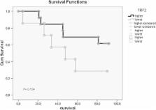
          Both five year overall- and disease-free survival rates were longer in patients with higher TRF2 expression compared to lower expression, but results were not statistically significant (69.2% vs 28.9%, respectively). Mean local recurrence-free survivals (LRF) were very close ( 58.6, CI: 44.3-72.9 vs 54.5, CI: 32.1-76.9 months) for high and low expressions, respectively. Cumulative proportion of LRF at the end of five year period was 76.9% for high and 57.1% for low TRF2 expression (P = 0.75). Statistically significant difference was found between survival ratios and Bcl-xL and p53 gene expressions, but not with TRF2. A respectable correlation between TRF2 expression and apoptosis along with distant metastasis was noted (P = 0.045 and 0.036, respectively). Additionally, high TRF2 expression levels had a positive impact in five year survival rate of stage IIIB-IVA patients (P = 0.04).

          Conclusions

          Our results support the role of TRF2 in apoptosis and imply a positive relation with distant metastases and survival in advanced stage patients. The remarkable difference in survival periods of patients with different TRF2 expressions suggest that TRF2 may be a candidate factor to estimate survival for cervical cancer, a preliminary observation which should further be verified with a larger cohort.

          Related collections

          Most cited references59

          • Record: found
          • Abstract: found
          • Article: not found

          DNA damage foci at dysfunctional telomeres.

          We report cytologic and genetic data indicating that telomere dysfunction induces a DNA damage response in mammalian cells. Dysfunctional, uncapped telomeres, created through inhibition of TRF2, became associated with DNA damage response factors, such as 53BP1, gamma-H2AX, Rad17, ATM, and Mre11. We refer to the domain of telomere-associated DNA damage factors as a Telomere Dysfunction-Induced Focus (TIF). The accumulation of 53BP1 on uncapped telomeres was reduced in the presence of the PI3 kinase inhibitors caffeine and wortmannin, which affect ATM, ATR, and DNA-PK. By contrast, Mre11 TIFs were resistant to caffeine, consistent with previous findings on the Mre11 response to ionizing radiation. A-T cells had a diminished 53BP1 TIF response, indicating that the ATM kinase is a major transducer of this pathway. However, in the absence of ATM, TRF2 inhibition still induced TIFs and senescence, pointing to a second ATM-independent pathway. We conclude that the cellular response to telomere dysfunction is governed by proteins that also control the DNA damage response. TIFs represent a new tool for evaluating telomere status in normal and malignant cells suspected of harboring dysfunctional telomeres. Furthermore, induction of TIFs through TRF2 inhibition provides an opportunity to study the DNA damage response within the context of well-defined, physically marked lesions.
            Bookmark
            • Record: found
            • Abstract: found
            • Article: not found

            Protection of mammalian telomeres.

            Telomeres allow cells to distinguish natural chromosome ends from damaged DNA. When telomere function is disrupted, a potentially lethal DNA damage response can ensue, DNA repair activities threaten the integrity of chromosome ends, and extensive genome instability can arise. It is not clear exactly how the structure of telomere ends differs from sites of DNA damage and how telomeres protect chromosome ends from DNA repair activities. What are the defining structural features of telomeres and through which mechanisms do they ensure chromosome end protection? What is the molecular basis of the telomeric cap and how does it act to sequester the chromosome end? Here I discuss data gathered in the last few years, suggesting that the protection of human chromosome ends primarily depends on the telomeric protein TRF2 and that telomere capping involves the formation of a higher order structure, the telomeric loop or t-loop.
              Bookmark
              • Record: found
              • Abstract: found
              • Article: not found

              Papillomavirus E6 and E7 proteins and their cellular targets.

              The mucosal human papillomaviruses (HPVs) infect human genital and oral epithelial cells and cause lesions ranging in severity from benign to malignant. HPV associated malignancies include cervical and other anogenital cancers as well as a subpopulation of head and neck cancers. Viral infection of epidermal stem or transit amplifying cells can result in long term viral persistence, and the development of carcinogenesis over a significant amount of time then requires additional cooperating genetic hits. Only the so-called high risk HPV types mediate human carcinogenesis, whereas the low risk HPVs have been linked to benign epithelial lesions that are not generally life threatening, but nonetheless are a major health burden. Expression of the high risk HPV E6 and E7 oncogenes is sufficient for primary human keratinocyte immortalization and is required for initiation and all subsequent stages of carcinogenic progression. Together with the finding that high levels of E6/E7 are a unifying hallmark of HPV positive cancers, these two genes are presumed to be the relevant virus-derived transformation stimuli in humans. E6 and E7 proteins do not possess intrinsic enzymatic activities, but instead function though a number of direct and indirect interactions with cellular proteins, a number of which are well known cellular tumor suppressors. We will summarize here current insights into E6 and E7 interactions with specific cellular targets that stimulate aspects of the viral life cycle, interfere with cell cycle controls and promote carcinogenic processes.
                Bookmark

                Author and article information

                Contributors
                drsevgi@hekim.net
                pinarmet@yahoo.com
                zerrinozgen34@hotmail.com
                hazanozyurt@yahoo.com
                nedimeserakinci@gmail.com
                oyaorun@yahoo.com
                Journal
                Biol Res
                Biol. Res
                Biological Research
                BioMed Central (London )
                0716-9760
                0717-6287
                25 November 2014
                2014
                : 47
                : 1
                : 61
                Affiliations
                [ ]Clinic of Radiation Oncology, Dr. Lutfi Kirdar Kartal Training and Research Hospital, Semsi Denizer Street, Istanbul, 34890 Turkey
                [ ]Biophysics Department, Marmara University School of Medicine, Maltepe Basibuyuk Yolu Street, Istanbul, 34854 Turkey
                [ ]Department of Radiation Oncology, Marmara University, School of Medicine, Muhsin Yazicioglu Street, Istanbul, 34890 Turkey
                [ ]Near East University, Faculty of Medicine, Medical Genetics, 922022 Lefkosa, KKTC Mersin 10, Turkey
                Article
                72
                10.1186/0717-6287-47-61
                4335779
                25654471
                7069a7e2-0fd6-4a0e-a38e-67076c4ae3d7
                © Ozden et al.; licensee BioMed Central Ltd. 2014

                This article is published under license to BioMed Central Ltd. This is an Open Access article distributed under the terms of the Creative Commons Attribution License ( http://creativecommons.org/licenses/by/4.0), which permits unrestricted use, distribution, and reproduction in any medium, provided the original work is properly credited. The Creative Commons Public Domain Dedication waiver ( http://creativecommons.org/publicdomain/zero/1.0/) applies to the data made available in this article, unless otherwise stated.

                History
                : 7 July 2014
                : 13 October 2014
                Categories
                Research Article
                Custom metadata
                © The Author(s) 2014

                cervical cancer,radiotherapy,telomere repeat-binding factors 2,b-cell lymphoma-extra large,apoptosis

                Comments

                Comment on this article