1
views
0
recommends
+1 Recommend
0 collections
    0
    shares
      • Record: found
      • Abstract: found
      • Article: found
      Is Open Access

      Asymptomatic Patient With Incarcerated Gravid Uterus Diagnosed in the Third Trimester: A Case Report of a Rare Potential Obstetric Emergency

      case-report

      Read this article at

      Bookmark
          There is no author summary for this article yet. Authors can add summaries to their articles on ScienceOpen to make them more accessible to a non-specialist audience.

          Abstract

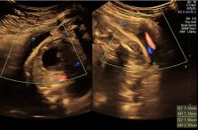
          Incarcerated gravid uterus (IGU) is a rare condition that occurs when a retropositioned gravid uterus becomes entrapped within the pelvic cavity. Most patients present around the 17th week of pregnancy with symptoms such as pelvic fullness, urinary incontinence, abdominal pain, constipation, and vaginal bleeding. Rarely, patients are asymptomatic throughout pregnancy, leaving IGU undiagnosed and untreated. Here, we present an asymptomatic 26-year-old female who presented at 30 weeks of gestation with severe intrauterine growth retardation (IUGR) on serial obstetric ultrasounds. Further evaluation with ultrasound and MRI revealed an incarcerated uterus. This was complicated by severe fetal IUGR, abnormal biophysical profile, and oligohydramnios. This case highlights the importance of early diagnosis and treatment of IGU in order to prevent complications associated with the condition. Clinicians should be aware that, although uncommon, patients with IGU may be asymptomatic and that diagnosis should depend primarily on imaging findings rather than symptoms.

          Related collections

          Most cited references6

          • Record: found
          • Abstract: found
          • Article: not found

          Recurrent incarceration and/or sacculation of the gravid uterus: a review.

          Retroverted uterine incarceration with sacculation of the anterior wall is reported to occur approximately in 1/3000 pregnancies. A literature search identified only one case report of incarceration of an anteflexed gravid uterus and six reported cases of recurrent incarceration and/or sacculation. We present a case of an incarceration of an anteflexed uterus in the first pregnancy, followed by a retroflexed incarceration in the second pregnancy. From this, a review is presented on recurrent uterine incarceration and/or sacculation.
            Bookmark
            • Record: found
            • Abstract: found
            • Article: not found

            Prenatal sonographic and MRI findings in a pregnancy complicated by uterine sacculation: case report and review of the literature.

            Approximately 15% of women have a retroverted uterus prior to pregnancy, and retroversion occurs in 11% of women in the first trimester of pregnancy. However, the uterus usually moves to an upward position before 14 weeks' gestation. Incarceration and sacculation of a retroverted uterus occur in 1 in 3000 pregnancies and are difficult to diagnose. They have often been missed until shortly before delivery and can lead to serious obstetric emergencies such as labor dystocia, uterine rupture, retained placenta and uncontrollable postpartum hemorrhage. Performing a Cesarean section without correct diagnosis may cause difficulties in identifying the bladder and the cervix, and therefore in opening the lower uterine segment. This leads to bladder injuries, vaginal transsection and trans- or supracervical hysterectomy. Early diagnosis and detailed scanning are crucial for the obstetric management and operative approach.We report a case of an incarcerated uterus in a patient presenting at 24 weeks' gestation with severe bilateral flank and lower abdominal pain. The symptoms were misdiagnosed as appendicitis. Digital examination revealed a ventralized vaginal axis. The cervix was not palpable. The clinical course, and two- and three-dimensional ultrasound and magnetic resonance imaging findings, are presented. The delivery was performed by midline laparotomy Cesarean section. The management for different gestational ages and a review of the literature are discussed.
              Bookmark
              • Record: found
              • Abstract: found
              • Article: not found

              The incarcerated uterus: a review of MRI and ultrasound imaging appearances.

              The objective of this article is to review the MRI and ultrasound appearances of incarcerated uterus.
                Bookmark

                Author and article information

                Journal
                Cureus
                Cureus
                2168-8184
                Cureus
                Cureus (Palo Alto (CA) )
                2168-8184
                12 September 2023
                September 2023
                : 15
                : 9
                : e45117
                Affiliations
                [1 ] Department of Radiology, Lake Erie College of Osteopathic Medicine, Erie, USA
                [2 ] Department of Radiology, University Hospitals Cleveland Medical Center/Case Western Reserve University, Cleveland, USA
                Author notes
                Article
                10.7759/cureus.45117
                10568245
                37842415
                7096daf2-ee8f-40b8-9e51-d6171e9f1d6e
                Copyright © 2023, Eskander et al.

                This is an open access article distributed under the terms of the Creative Commons Attribution License, which permits unrestricted use, distribution, and reproduction in any medium, provided the original author and source are credited.

                History
                : 12 September 2023
                Categories
                Obstetrics/Gynecology
                Radiology
                General Surgery

                classical cesarean,pelvic mri,pelvic ultrasound,oligohydramnios,cesarean section,pregnancy,obstetrics and gynecology,intrauterine growth retardation,incarcerated gravid uterus,incarcerated uterus

                Comments

                Comment on this article