8
views
0
recommends
+1 Recommend
0 collections
    0
    shares
      • Record: found
      • Abstract: found
      • Article: found
      Is Open Access

      Arthroscopically assisted fixation of the lesser trochanter fracture: a case series

      research-article

      Read this article at

      Bookmark
          There is no author summary for this article yet. Authors can add summaries to their articles on ScienceOpen to make them more accessible to a non-specialist audience.

          Abstract

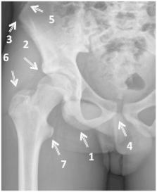
          Avulsion fractures of the lesser trochanter in adolescents are uncommon. This injury is a result of a sudden forceful contraction of the iliopsoas tendon. It usually occurs during vigorous sport activity. Historically, these injuries were treated non-operatively, with guarded results, including weak hip flexor strength and non-union, hindering return to competitive sport. We report a series of three arthroscopically assisted fracture fixations performed by the senior author, using cannulated screw fixation in two cases and an anchor in one case. Mobilization was commenced immediately following surgery, allowing weight bearing as tolerated using crutches for 4 weeks, thereafter unaided walking was allowed. Patients were assessed at 2 weeks, 6 weeks, 3 months and 1-year post-operatively. Radiographs were utilized to confirm full union. All three patients were able to mobilize unaided by 4 weeks post-operatively and two of the three patients returned to competitive sport at 3 months. Near—anatomical union was achieved in all cases. No complications were noted during surgery and the peri-operative period in our series. The utilization of arthroscopic reduction and fixation of avulsion of the lesser trochanter results in good fixation and allows a faster recovery with a return to sports activity, and therefore, we suggest it as a viable treatment option for such injuries.

          Related collections

          Most cited references35

          • Record: found
          • Abstract: found
          • Article: not found

          Acute avulsion fractures of the pelvis in adolescent competitive athletes: prevalence, location and sports distribution of 203 cases collected.

          To describe the prevalence, location and sports distribution of pelvic avulsion fractures in adolescent competitive athletes. One thousand two hundred and thirty-eight radiographs of the pelvis taken for focal traumatic symptoms in athletes with an age range of 11-35 years over a period of 22 years were reviewed. One hundred and ninety-eight adolescent athletes were affected by 203 avulsion fractures of the pelvic apophyses (five cases presented multiple locations). The localisation was the ischial tuberosity (IT) in 109 cases, anterior inferior iliac spine (AIIS) in 45 cases, anterior superior iliac spine (ASIS) in 39 cases, superior corner of pubic symphysis (SCPS) in 7 cases and iliac crest (IC) in 3 cases. Soccer (74 cases) and gymnastics (55 cases) were the sports with the highest number of avulsion fractures documented. Apophyseal avulsion fractures of the pelvis in adolescent competitive athletes are most common in soccer and gymnastics. The lesions are usually the consequence of sudden and forceful muscle-tendon contractions during sport activities. Plain radiographs, are determinant for the diagnosis.
            Bookmark
            • Record: found
            • Abstract: found
            • Article: not found

            Ischiofemoral impingement.

            Femoroacetabular impingement is a well-documented cause of hip pain. There is, however, increasing evidence for the presence of a previously unrecognised impingement-type condition around the hip - ischiofemoral impingement. This is caused by abnormal contact between the lesser trochanter of the femur and the ischium, and presents as atypical groin and/or posterior buttock pain. The symptoms are gradual in onset and may be similar to those of iliopsoas tendonitis, hamstring injury or bursitis. The presence of ischiofemoral impingement may be indicated by pain caused by a combination of hip extension, adduction and external rotation. Magnetic resonance imaging demonstrates inflammation and oedema in the ischiofemoral space and quadratus femoris, and is distinct from an acute tear. To date this has only appeared in the specialist orthopaedic literature as a problem that has developed after total hip replacement, not in the unreplaced joint.
              Bookmark
              • Record: found
              • Abstract: found
              • Article: not found

              Avulsion injuries of the pelvis and proximal femur.

              Twenty cases of avulsion fractures of the apophyses of the pelvis and proximal femur were reviewed. Most occurred in male adolescents engaged in active sports. Pain, often with little external evidence of trauma, was the most frequent presenting symptom. Conservative, nonsurgical treatment was successful in all the patients. The radiographic appearance of each type of apophyseal injury is demonstrated. Recognition of the initial deformity and of the patterns of repair is important to avoid unnecessary evaluation (tomography, radionuclide scans) and inappropriate therapy. While productive changes with healing are most likely to occur in the region of the ischium, any of the other sites may heal with abundant bone and may mimic neoplasm. In the proper clinical setting, bony changes at these sites should prompt the radiologist to consider a traumatic etiology.
                Bookmark

                Author and article information

                Journal
                J Hip Preserv Surg
                J Hip Preserv Surg
                jhps
                jhps
                Journal of Hip Preservation Surgery
                Oxford University Press
                2054-8397
                July 2014
                22 August 2014
                : 1
                : 1
                : 27-32
                Affiliations
                1. Norwest Private Hospital, Bella Vista 2153, Sydney, Australia 2. Department of Joint Arthroplasty and Clinical Science, Mare Clinic, Universitatsklinikum Schleswig-Holstein, Kiel, Germany 3. University Of Notre Dame, Australia School of Medicine, Sydney, Australia
                Author notes
                *Correspondence author: A. Khemka. E-mail: adk_sun@ 123456hotmail.com
                Article
                hnu006
                10.1093/jhps/hnu006
                4765264
                27011799
                743c1f4f-63a6-45e8-9685-bca9bf3f1fb4
                © The Author 2014. Published by Oxford University Press.

                This is an Open Access article distributed under the terms of the Creative Commons Attribution License ( http://creativecommons.org/licenses/by/4.0/), which permits unrestricted reuse, distribution, and reproduction in any medium, provided the original work is properly cited.

                History
                : 29 April 2014
                : 13 June 2014
                : 13 July 2014
                Page count
                Pages: 6
                Categories
                Research Articles

                Comments

                Comment on this article