12
views
0
recommends
+1 Recommend
0 collections
    0
    shares
      • Record: found
      • Abstract: found
      • Article: found
      Is Open Access

      Lupus antibodies induce behavioral changes mediated by microglia and blocked by ACE inhibitors

      research-article

      Read this article at

      Bookmark
          There is no author summary for this article yet. Authors can add summaries to their articles on ScienceOpen to make them more accessible to a non-specialist audience.

          Abstract

          Nestor et al. examine how lupus antibodies that enter the brain cause neuronal dysfunction and cognitive impairment. The results show that activated microglia are critical for neuronal damage and that inhibiting them can preserve neuronal function and cognition.

          Abstract

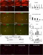
          Cognitive impairment occurs in 40–90% of patients with systemic lupus erythematosus (SLE), which is characterized by autoantibodies to nuclear antigens, especially DNA. We discovered that a subset of anti-DNA antibodies, termed DNRAbs, cross reacts with the N-methyl- d-aspartate receptor (NMDAR) and enhances NMDAR signaling. In patients, DNRAb presence associates with spatial memory impairment. In a mouse model, DNRAb-mediated brain pathology proceeds through an acute phase of excitotoxic neuron loss, followed by persistent alteration in neuronal integrity and spatial memory impairment. The latter pathology becomes evident only after DNRAbs are no longer detectable in the brain. Here we investigate the mechanism of long-term neuronal dysfunction mediated by transient exposure to antibody. We show that activated microglia and C1q are critical mediators of neuronal damage. We further show that centrally acting inhibitors of angiotensin-converting enzyme (ACE) can prevent microglial activation and preserve neuronal function and cognitive performance. Thus, ACE inhibition represents a strong candidate for clinical trials aimed at mitigating cognitive dysfunction.

          Related collections

          Most cited references36

          • Record: found
          • Abstract: found
          • Article: not found

          Novel role of PKR in inflammasome activation and HMGB1 release

          The inflammasome regulates release of caspase activation-dependent cytokines, including IL-1β, IL-18, and high-mobility group box 1 (HMGB1) 1-5 . During the course of studying HMGB1 release mechanisms, we discovered an important role of double-stranded RNA dependent protein kinase (PKR) in inflammasome activation. Exposure of macrophages to inflammasome agonists induced PKR autophosphorylation. PKR inactivation by genetic deletion or pharmacological inhibition severely impaired inflammasome activation in response to double-stranded RNA, ATP, monosodium urate, adjuvant aluminum, rotenone, live E. coli, anthrax lethal toxin, DNA transfection, and S. Typhimurium infection. PKR deficiency significantly inhibited the secretion of IL-1beta, IL-18 and HMGB1 in E. coli-induced peritonitis. PKR physically interacts with multiple inflammasome components, including NLR family pyrin domain-containing 3 (NLRP3), NLR family pyrin domain-containing 1 (NLRP1), NLR family CARD domain-containing protein 4 (NLRC4), Absent in melanoma 2 (AIM2), and broadly regulates inflammasome activation. PKR autophosphorylation in a cell free system with recombinant NLRP3, ASC and pro-casapse-1 reconstitutes inflammasome activity. These results reveal a critical role of PKR in inflammasome activation, and indicate that it should be possible to pharmacologically target this molecule to treat inflammation.
            Bookmark
            • Record: found
            • Abstract: found
            • Article: found
            Is Open Access

            Cell-specific deletion of C1qa identifies microglia as the dominant source of C1q in mouse brain

            Background The complement cascade not only provides protection from infection but can also mediate destructive inflammation. Complement is also involved in elimination of neuronal synapses which is essential for proper development, but can be detrimental during aging and disease. C1q, required for several of these complement-mediated activities, is present in the neuropil, microglia, and a subset of interneurons in the brain. Methods To identify the source(s) of C1q in the brain, the C1qa gene was selectively inactivated in the microglia or Thy-1+ neurons in both wild type mice and a mouse model of Alzheimer’s disease (AD), and C1q synthesis assessed by immunohistochemistry, QPCR, and western blot analysis. Results While C1q expression in the brain was unaffected after inactivation of C1qa in Thy-1+ neurons, the brains of C1qa FL/FL :Cx3cr1 CreERT2 mice in which C1qa was ablated in microglia were devoid of C1q with the exception of limited C1q in subsets of interneurons. Surprisingly, this loss of C1q occurred even in the absence of tamoxifen by 1 month of age, demonstrating that Cre activity is tamoxifen-independent in microglia in Cx3cr1 CreERT2/WganJ mice. C1q expression in C1qa FL/FL : Cx3cr1 CreERT2/WganJ mice continued to decline and remained almost completely absent through aging and in AD model mice. No difference in C1q was detected in the liver or kidney from C1qa FL/FL : Cx3cr1 CreERT2/WganJ mice relative to controls, and C1qa FL/FL : Cx3cr1 CreERT2/WganJ mice had minimal, if any, reduction in plasma C1q. Conclusions Thus, microglia, but not neurons or peripheral sources, are the dominant source of C1q in the brain. While demonstrating that the Cx3cr1 CreERT2/WganJ deleter cannot be used for adult-induced deletion of genes in microglia, the model described here enables further investigation of physiological roles of C1q in the brain and identification of therapeutic targets for the selective control of complement-mediated activities contributing to neurodegenerative disorders. Electronic supplementary material The online version of this article (doi:10.1186/s12974-017-0814-9) contains supplementary material, which is available to authorized users.
              Bookmark
              • Record: found
              • Abstract: found
              • Article: not found

              Microglial repopulation resolves inflammation and promotes brain recovery after injury.

              Microglia mediate chronic neuroinflammation following central nervous system (CNS) disease or injury, and in doing so, damage the local brain environment by impairing recovery and contributing to disease processes. Microglia are critically dependent on signaling through the colony-stimulating factor 1 receptor (CSF1R) and can be eliminated via administration of CSF1R inhibitors. Resolving chronic neuroinflammation represents a universal goal for CNS disorders, but long-term microglial elimination may not be amenable to clinical use. Notably, withdrawal of CSF1R inhibitors stimulates new microglia to fully repopulate the CNS, affording an opportunity to renew this cellular compartment. To that end, we have explored the effects of acute microglial elimination, followed by microglial repopulation, in a mouse model of extensive neuronal loss. Neuronal loss leads to a prolonged neuroinflammatory response, characterized by the presence of swollen microglia expressing CD68 and CD45, as well as elevated levels of cytokines, chemokines, complement, and other inflammatory signals. These collective responses are largely resolved by microglial repopulation. Furthermore, microglial repopulation promotes functional recovery in mice, with elevated plus maze performance matching that of uninjured mice, despite the loss of 80% of hippocampal neurons. Analyses of synaptic surrogates revealed increases in PSD95 and synaptophysin puncta with microglial repopulation, suggesting that these cells sculpt and regulate the synaptic landscape. Thus, our results show that short-term microglial elimination followed by repopulation may represent a clinically feasible and novel approach to resolve neuroinflammatory events and promote brain recovery.
                Bookmark

                Author and article information

                Journal
                J Exp Med
                J. Exp. Med
                jem
                jem
                The Journal of Experimental Medicine
                Rockefeller University Press
                0022-1007
                1540-9538
                01 October 2018
                : 215
                : 10
                : 2554-2566
                Affiliations
                [1 ]Center for Autoimmune Musculoskeletal and Hematopoietic Diseases, The Feinstein Institute for Medical Research, Manhasset, NY
                [2 ]Donald & Barbara Zucker School of Medicine at Hofstra/Northwell, Manhasset, NY
                [3 ]Rheumatology and Infectious Diseases, Kitasato University School of Medicine, Kanagawa, Japan
                [4 ]Department of Rheumatology, Endocrinology and Nephrology Faculty of Medicine and Graduate School of Medicine Hokkaido University, Sapporo, Japan
                [5 ]Boston Children’s Hospital and Department of Pediatrics, Harvard Medical School, Boston, MA
                [6 ]Department of Pathology and Laboratory Medicine, Lewis Katz School of Medicine, Temple University, Philadelphia, PA
                [7 ]Kirby Neurobiology Center Boston Children's Hospital, Boston, MA
                [8 ]Center for Biomedical Sciences, The Feinstein Institute for Medical Research, Manhasset, NY
                Author notes
                Correspondence to Betty Diamond: bdiamond@ 123456northwell.edu
                [*]

                J. Nestor and Y. Arinuma contributed equally to this paper.

                Author information
                http://orcid.org/0000-0002-3250-3804
                Article
                20180776
                10.1084/jem.20180776
                6170183
                30185634
                751a32dc-e2eb-40e7-8124-785bea1db5f6
                © 2018 Nestor et al.

                This article is distributed under the terms of an Attribution–Noncommercial–Share Alike–No Mirror Sites license for the first six months after the publication date (see http://www.rupress.org/terms/). After six months it is available under a Creative Commons License (Attribution–Noncommercial–Share Alike 4.0 International license, as described at https://creativecommons.org/licenses/by-nc-sa/4.0/).

                History
                : 25 April 2018
                : 06 July 2018
                : 02 August 2018
                Funding
                Funded by: National Institutes of Health, DOI http://dx.doi.org/10.13039/100000002;
                Award ID: NIH P01AI073693
                Award ID: NIH R03-AR065157
                Categories
                Research Articles
                Article
                300

                Medicine
                Medicine

                Comments

                Comment on this article