125
views
0
recommends
+1 Recommend
0 collections
    30
    shares
      • Record: found
      • Abstract: found
      • Article: found
      Is Open Access

      Lumbar Spinal Stenosis Due to a Large Calcified Mass in the Ligamentum Flavum

      case-report

      Read this article at

      Bookmark
          There is no author summary for this article yet. Authors can add summaries to their articles on ScienceOpen to make them more accessible to a non-specialist audience.

          Abstract

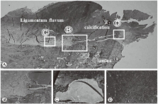
          We describe a rare case of lumbar spinal stenosis due to a large calcified mass in the ligamentum flavum. This patient presented with a 12-month history of severe right leg pain and intermittent claudication. A computed tomography scan was performed, revealing a large calcified mass on the ligamentum flavum at the right-hand side of the lumbar spinal canal. We performed a laminotomy at the L4/5 level with resection of the calcified mass from the ligamentum flavum. The findings of various analyses suggested that the calcified mass consisted mostly of Ca 3(PO 4) 2 and calcium phosphate intermixed with protein and water. The calcified mass in the ligamentum flavum was causing lumbar spinal stenosis. Surgical decompression by resection of the mass was effective in this patient. The calcified material was composed mainly of elements derived from calcium phosphate. Degenerative changes in the ligamentum flavum of the lumbar spine may have been involved in the production of this calcified mass.

          Related collections

          Most cited references16

          • Record: found
          • Abstract: found
          • Article: not found

          The pathology of ligamentum flavum in degenerative lumbar disease.

          A pathologic study of the ligamentum flavum in degenerative lumbar disease. To elucidate the clinical significance of each pathologic finding of the ligamentum flavum. In many reports, researchers observed the ligamentum flavum removed partially during surgery and did not evaluate the whole image of the ligamentum flavum. In addition, there are only a few reports that examined the possible association between various histologic findings and clinical findings. And, thus, there are many unclear points in the clinical significance indicated by each pathologic finding. The study participants were 50 patients with degenerative lumbar diseases who underwent surgical decompression with removal of the ligamentum flavum of the affected spinal level. Tissue specimens of the removed ligamentum flavum in cross section were prepared, and changes in the elastic fibers and collagen fibers were evaluated in three grades to evaluate the whole image. In addition, we observed the presence or absence of any focal lesions and statistically analyzed the possible association between these histologic findings and clinical symptoms or image findings. In regard to the association between histologic findings and clinical symptoms or image findings, calcification was observed in significantly older patients, who tended to have low scores in preoperative JOA score, and was frequently observed in patients with cauda equina symptoms. Patients with ossification had a significantly greater % slip, and chondroid cells were frequently observed in patients with spondylolisthesis. Various pathologic findings provided important foundations for discussing the pathogenesis of lesions in ligamentum flavum. Calcification was frequently observed in elderly patients and those with cauda equina symptoms, and these patients tended to have severer preoperative symptoms. Chondroid cells were frequently observed in patients with spondylolisthesis, and patients with ossification had a greater % slip, suggesting involvement of mechanical load in ossification of ligaments. The pathologic findings were significantly related to the clinical features, and these findings will be profitable for understanding the pathogenesis of degenerative lumbar disease.
            Bookmark
            • Record: found
            • Abstract: found
            • Article: not found

            Histology of the ligamentum flavum in patients with degenerative lumbar spinal stenosis.

            The degree of calcification as well as the structural changes of the elastic fibres in the ligamentum flavum in patients with degenerative lumbar spinal stenosis were evaluated and the results were compared to those of patients without spinal stenosis. In 21 patients (13 male, 8 female) with lumbar spinal stenosis the ligamentum flavum was removed, histologically processed and stained. The calcification, the elastic/collagenous fibre ratio as well as the configuration of the fibres were evaluated with an image analyzing computer. As a control group, 20 ligaments of 10 human corpses were processed in the same way. The results were statistically analysed using the Mann-Whitney-Wilcoxon test (alpha = 0.05) and the t-test (alpha = 0.05). Nearly all the ligaments of patients with lumbar spinal stenosis were calcified (average 0.17%, maximum 3.8%) and showed relevant fibrosis with decreased elastic/collagenous fibre ratio. There was a significant correlation between age and histological changes (P<0.05). In the control group we only found minimal calcification in 3 of 20 segments (average 0.015%). No relevant fibrosis was found and the configuration of elastic fibres showed no pathologic changes. The results of this study illustrate the important role of histological changes of the ligamentum flavum for the aetiology of lumbar spinal stenosis.
              Bookmark
              • Record: found
              • Abstract: found
              • Article: not found

              Calcium pyrophosphate dehydrate crystal deposition in the ligamentum flavum of the cervical spine: histopathological and immunohistochemical findings.

              To investigate the histopathological and immunohistochemical properties of degenerative changes in the ligamentum flavum of the cervical spine with calcium crystal deposition. Sections of the calcified ligamentum flavum harvested from 26 patients who required cervical decompression were examined by scanning electron microscopy (SEM), energy dispersive X-ray microanalysis, immunohistochemical staining [for transforming growth factor (TGF)-Beta, vascular endothelial growth factor (VEGF), Sox9, and Msx2] and terminal deoxynucleotidyl transferase (TdT)-mediated dUTP-biotin nick end labelling (TUNEL) method (for cell apoptosis). Energy dispersive x-ray microanalysis and SEM confirmed the deposited calcium to be calcium pyrophosphate dihydrate (CPPD) crystals. The calcified ligamentum flavum showed disorganisation of the elastic fibre bundles together with increased collagen fibrils in the matrix. Abundant hypertrophic chondrocytes were noted around the calcified lesions, which were strongly immunoreactive to TGF-Beta and VEGF. Staining for Sox9 was positive in metaplastic chondrocytes but negative in hypertrophic chondrocytes. Both chondrocytes and mesenchymal cells were positive for Msx2. TUNEL-positive hypertrophic chondrocytes were significantly more noticeable in nodular than diffusely scattered type of CPPD deposition. Calcium crystal deposition in the cervical ligamentum flavum seems to progress with reduction in elastic fibres, increase in collagen fibrils in the matrix, and migration of metaplastic hypertrophic chondrocytes, whose differentiation is controlled by cytokines and transcriptional factors, and potentially regulate crystal formation. The presence of abundant TUNEL-positive hypertrophic chondrocytes around CPPD deposition suggests that materials from apoptotic cells play some role in crystal deposition.
                Bookmark

                Author and article information

                Journal
                Asian Spine J
                Asian Spine J
                ASJ
                Asian Spine Journal
                Korean Society of Spine Surgery
                1976-1902
                1976-7846
                September 2013
                04 September 2013
                : 7
                : 3
                : 236-241
                Affiliations
                [1 ]Department of Orthopaedic Surgery, Faculty of Medicine, University of Toyama, Toyama, Japan.
                [2 ]Department of Orthopaedic Surgery, Machida Municipal Hospital, Tokyo, Japan.
                [3 ]Department of Pathology, Niigata Prefectural Central Hospital, Niigata, Japan.
                Author notes
                Corresponding author: Shoji Seki. Department of Orthopaedic Surgery, Faculty of Medicine, University of Toyama, 2630 Sugitani, Toyama 930-0194, Japan. Tel: +81-76-434-7353, Fax: +81-76-434-5035, seki@ 123456med.u-toyama.ac.jp
                Article
                10.4184/asj.2013.7.3.236
                3779778
                24066222
                7673c00d-f16c-48e2-903c-37c659a1b54e
                Copyright © 2013 by Korean Society of Spine Surgery

                This is an Open Access article distributed under the terms of the Creative Commons Attribution Non-Commercial License ( http://creativecommons.org/licenses/by-nc/3.0/) which permits unrestricted non-commercial use, distribution, and reproduction in any medium, provided the original work is properly cited.

                History
                : 02 February 2012
                : 03 April 2012
                : 09 April 2012
                Categories
                Case Report

                Orthopedics
                spinal stenosis,calcification,ligamentum flavum,calcium phosphate
                Orthopedics
                spinal stenosis, calcification, ligamentum flavum, calcium phosphate

                Comments

                Comment on this article