Inviting an author to review:
Find an author and click ‘Invite to review selected article’ near their name.
Search for authorsSearch for similar articles
4
views
0
recommends
+1 Recommend
0 collections
    0
    shares
      • Record: found
      • Abstract: found
      • Article: found
      Is Open Access

      Human umbilical cord mesenchymal stromal cells-derived extracellular vesicles exert potent bone protective effects by CLEC11A-mediated regulation of bone metabolism

      research-article

      Read this article at

      Bookmark
          There is no author summary for this article yet. Authors can add summaries to their articles on ScienceOpen to make them more accessible to a non-specialist audience.

          Abstract

          Osteoporosis and osteoporotic fractures severely compromise quality of life in elderly people and lead to early death. Human umbilical cord mesenchymal stromal cell (MSC)-derived extracellular vesicles (hucMSC-EVs) possess considerable therapeutic effects in tissue repair and regeneration. Thus, in the present study, we investigated the effects of hucMSC-EVs on primary and secondary osteoporosis and explored the underlying mechanisms.

          Methods: hucMSCs were isolated and cultured. EVs were obtained from the conditioned medium of hucMSCs and determined by using transmission electron microscopy, dynamic light scattering and Western Blot analyses. The effects of hucMSC-EVs on ovariectomy-induced postmenopausal osteoporosis and tail suspension-induced hindlimb disuse osteoporosis in mouse models were assessed by using microcomputed tomography, biomechanical, histochemical and immunohistochemical, as well as histomorphometric analyses. Proteomic analysis was applied between hucMSC-EVs and hucMSCs to screen the candidate proteins that mediate hucMSC-EVs function. The effects of hucMSC-EVs on osteogenic and adipogenic differentiation of bone marrow mesenchymal stromal cells (BMSCs), and osteoclastogenesis of the macrophage cell line RAW264.7 in vitro were determined by using cytochemical staining and quantitative real-time PCR analysis. Subsequently, the roles of the key protein in hucMSC-EVs-induced regulation on BMSCs and RAW264.7 cells were evaluated.

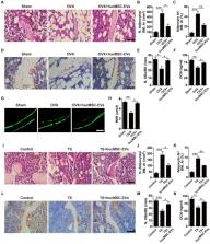
          Results: hucMSCs were able to differentiate into osteoblasts, adipocytes or chondrocytes and positively expressed CD29, CD44, CD73 and CD90, but negatively expressed CD34 and CD45. The morphological assessment revealed the typical cup- or sphere-shaped morphology of hucMSC-EVs with diameters predominantly ranging from 60 nm to 150 nm and expressed CD9, CD63, CD81 and TSG101. The systemic administration of hucMSC-EVs prevented bone loss and maintained bone strength in osteoporotic mice by enhancing bone formation, reducing marrow fat accumulation and decreasing bone resorption. Proteomic analysis showed that the potently pro-osteogenic protein, CLEC11A (C-type lectin domain family 11, member A) was very highly enriched in hucMSC-EVs. In addition, hucMSC-EVs enhanced the shift from adipogenic to osteogenic differentiation of BMSCs via delivering CLEC11A in vitro. Moreover, CLEC11A was required for the inhibitory effects of hucMSC-EVs on osteoclast formation.

          Conclusion: Our results suggest that hucMSC-EVs serve as a critical regulator of bone metabolism by transferring CLEC11A and may represent a potential agent for prevention and treatment of osteoporosis.

          Related collections

          Most cited references37

          • Record: found
          • Abstract: found
          • Article: not found

          Exosomes derived from miR-140-5p-overexpressing human synovial mesenchymal stem cells enhance cartilage tissue regeneration and prevent osteoarthritis of the knee in a rat model

          OBJECTIVES: Osteoarthritis (OA) is the most common joint disease throughout the world. Exosomes derived from miR-140-5p-overexpressing synovial mesenchymal stem cells (SMSC-140s) may be effective in treating OA. We hypothesized that exosomes derived from SMSC-140 (SMSC-140-Exos) would enhance the proliferation and migration abilities of articular chondrocytes (ACs) without harming extracellular matrix (ECM) secretion. METHODS: SMSCs were transfected with or without miR-140-5p. Exosomes derived from SMSCs or SMSC-140s (SMSC-Exos or SMSC-140-Exos) were isolated and identified. Proliferation, migration and ECM secretion were measured in vitro and compared between groups. The mechanism involving alternative Wnt signalling and activation of Yes-associated protein (YAP) was investigated using lentivirus, oligonucleotides or chemical drugs. The preventative effect of exosomes in vivo was measured using Safranin-O and Fast green staining and immunohistochemical staining. RESULTS: Wnt5a and Wnt5b carried by exosomes activated YAP via the alternative Wnt signalling pathway and enhanced proliferation and migration of chondrocytes with the side-effect of significantly decreasing ECM secretion. Highly-expressed miR-140-5p blocked this side-effect via RalA. SMSC-140-Exos enhanced the proliferation and migration of ACs without damaging ECM secretion in vitro, while in vivo, SMSC-140-Exos successfully prevented OA in a rat model. CONCLUSIONS: These findings highlight the promising potential of SMSC-140-Exos in preventing OA. We first found a potential source of exosomes and studied their merits and shortcomings. Based on our understanding of the molecular mechanism, we overcame the shortcomings by modifying the exosomes. Such exosomes derived from modified cells hold potential as future therapeutic strategies.
            Bookmark
            • Record: found
            • Abstract: found
            • Article: not found

            MicroRNA-188 regulates age-related switch between osteoblast and adipocyte differentiation.

            Bone marrow mesenchymal stem cells (BMSCs) exhibit an age-dependent reduction in osteogenesis that is accompanied by an increased propensity toward adipocyte differentiation. This switch increases adipocyte numbers and decreases the number of osteoblasts, contributing to age-related bone loss. Here, we found that the level of microRNA-188 (miR-188) is markedly higher in BMSCs from aged compared with young mice and humans. Compared with control mice, animals lacking miR-188 showed a substantial reduction of age-associated bone loss and fat accumulation in bone marrow. Conversely, mice with transgenic overexpression of miR-188 in osterix+ osteoprogenitors had greater age-associated bone loss and fat accumulation in bone marrow relative to WT mice. Moreover, using an aptamer delivery system, we found that BMSC-specific overexpression of miR-188 in mice reduced bone formation and increased bone marrow fat accumulation. We identified histone deacetylase 9 (HDAC9) and RPTOR-independent companion of MTOR complex 2 (RICTOR) as the direct targets of miR-188. Notably, BMSC-specific inhibition of miR-188 by intra-bone marrow injection of aptamer-antagomiR-188 increased bone formation and decreased bone marrow fat accumulation in aged mice. Together, our results indicate that miR-188 is a key regulator of the age-related switch between osteogenesis and adipogenesis of BMSCs and may represent a potential therapeutic target for age-related bone loss.
              Bookmark
              • Record: found
              • Abstract: found
              • Article: found
              Is Open Access

              Exosomes released by human umbilical cord mesenchymal stem cells protect against cisplatin-induced renal oxidative stress and apoptosis in vivo and in vitro

              Introduction Administration of bone marrow mesenchymal stem cells (MSCs) or secreted microvesicles improves recovery from acute kidney injury (AKI). However, the potential roles and mechanisms are not well understood. In the current study, we focused on the protective effect of exosomes derived from human umbilical cord mesenchymal stem cells (hucMSC-ex) on cisplatin-induced nephrotoxicity in vivo and in vitro. Methods We constructed cisplatin-induced AKI rat models. At 24 h after treatment with cisplatin, hucMSC-ex were injected into the kidneys via the renal capsule; human lung fibroblast (HFL-1)-secreted exosomes (HFL-1-ex) were used as controls. All animals were killed at day 5 after administration of cisplatin. Renal function, histological changes, tubular apoptosis and proliferation, and degree of oxidative stress were evaluated. In vitro, rat renal tubular epithelial (NRK-52E) cells were treated with or without cisplatin and after 6 h treated with or without exosomes. Cells continued to be cultured for 24 h, and were then harvested for western blotting, apoptosis and detection of degree of oxidative stress. Results After administration of cisplatin, there was an increase in blood urea nitrogen (BUN) and creatinine (Cr) levels, apoptosis, necrosis of proximal kidney tubules and formation of abundant tubular protein casts and oxidative stress in rats. Cisplatin-induced AKI rats treated with hucMSC-ex, however, showed a significant reduction in all the above indexes. In vitro, treatment with cisplatin alone in NRK-52E cells resulted in an increase in the number of apoptotic cells, oxidative stress and activation of the p38 mitogen-activated protein kinase (p38MAPK) pathway followed by a rise in the expression of caspase 3, and a decrease in cell multiplication, while those results were reversed in the hucMSCs-ex-treated group. Furthermore, it was observed that hucMSC-ex promoted cell proliferation by activation of the extracellular-signal-regulated kinase (ERK)1/2 pathway. Conclusions The results in the present study indicate that hucMSC-ex can repair cisplatin-induced AKI in rats and NRK-52E cell injury by ameliorating oxidative stress and cell apoptosis, promoting cell proliferation in vivo and in vitro. This suggests that hucMSC-ex could be exploited as a potential therapeutic tool in cisplatin-induced nephrotoxicity.
                Bookmark

                Author and article information

                Journal
                Theranostics
                Theranostics
                thno
                Theranostics
                Ivyspring International Publisher (Sydney )
                1838-7640
                2020
                16 January 2020
                : 10
                : 5
                : 2293-2308
                Affiliations
                [1 ]Department of Orthopedics, Xiangya Hospital, Central South University, Changsha, Hunan 410008, China.
                [2 ]Movement System Injury and Repair Research Center, Xiangya Hospital, Central South University, Changsha, Hunan 410008, China.
                [3 ]Department of Metabolism and Endocrinology, The First Affiliated Hospital, University of South China, Hengyang, Hunan 421001, China.
                [4 ]Department of Sports Medicine, Xiangya Hospital, Central South University, Changsha, Hunan 410008, China.
                [5 ]Xiangya Nursing School, Central South University, Changsha, Hunan 410013, China.
                [6 ]Department of Forensic Science, School of Basic Medical Science, Central South University, Changsha, Hunan 410013, China.
                [7 ]Hunan Key Laboratory of Organ Injury, Aging and Regenerative Medicine, Changsha, Hunan 410008, China.
                [8 ]Hunan Key Laboratory of Bone Joint Degeneration and Injury, Changsha, Hunan 410008, China.
                [9 ]National Clinical Research Center for Geriatric Disorders, Xiangya Hospital, Central South University, Changsha, Hunan 410008, China.
                Author notes
                ✉ Corresponding author: Hui Xie, Ph.D., Department of Orthopedics, Xiangya Hospital, Central South University, #87 Xiangya Road, Changsha, Hunan 410008, China. Tel: 86-0731-84327068; E-mail: huixie@ 123456csu.edu.cn

                *Yin Hu and Yan Zhang contributed equally to this work.

                Competing Interests: The authors have declared that no competing interest exists.

                Article
                thnov10p2293
                10.7150/thno.39238
                7019162
                32089743
                775e6ec8-ce32-4ee2-9365-9c12c0fad32d
                © The author(s)

                This is an open access article distributed under the terms of the Creative Commons Attribution License ( https://creativecommons.org/licenses/by/4.0/). See http://ivyspring.com/terms for full terms and conditions.

                History
                : 12 August 2019
                : 30 November 2019
                Categories
                Research Paper

                Molecular medicine
                extracellular vesicles,umbilical cord-derived mesenchymal stromal cells,bone homeostasis,osteoporosis,clec11a

                Comments

                Comment on this article