41
views
0
recommends
+1 Recommend
0 collections
    0
    shares
      • Record: found
      • Abstract: found
      • Article: found
      Is Open Access

      ALS/FTD‐associated FUS activates GSK‐3β to disrupt the VAPB–PTPIP51 interaction and ER–mitochondria associations

      research-article

      Read this article at

      Bookmark
          There is no author summary for this article yet. Authors can add summaries to their articles on ScienceOpen to make them more accessible to a non-specialist audience.

          Abstract

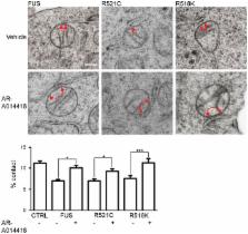
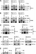
          Defective FUS metabolism is strongly associated with amyotrophic lateral sclerosis and frontotemporal dementia ( ALS/ FTD), but the mechanisms linking FUS to disease are not properly understood. However, many of the functions disrupted in ALS/ FTD are regulated by signalling between the endoplasmic reticulum ( ER) and mitochondria. This signalling is facilitated by close physical associations between the two organelles that are mediated by binding of the integral ER protein VAPB to the outer mitochondrial membrane protein PTPIP51, which act as molecular scaffolds to tether the two organelles. Here, we show that FUS disrupts the VAPBPTPIP51 interaction and ER–mitochondria associations. These disruptions are accompanied by perturbation of Ca 2+ uptake by mitochondria following its release from ER stores, which is a physiological read‐out of ER–mitochondria contacts. We also demonstrate that mitochondrial ATP production is impaired in FUS‐expressing cells; mitochondrial ATP production is linked to Ca 2+ levels. Finally, we demonstrate that the FUS‐induced reductions to ER–mitochondria associations and are linked to activation of glycogen synthase kinase‐3β ( GSK‐3β), a kinase already strongly associated with ALS/ FTD.

          Related collections

          Most cited references22

          • Record: found
          • Abstract: found
          • Article: not found

          A mutation in sigma-1 receptor causes juvenile amyotrophic lateral sclerosis.

          Amyotrophic lateral sclerosis (ALS) is a neurodegenerative disorder characterized by loss of motor neurons in the brain and spinal cord, leading to muscle weakness and eventually death from respiratory failure. ALS is familial in about 10% of cases, with SOD1 mutations accounting for 20% of familial cases. Here we describe a consanguineous family segregating juvenile ALS in an autosomal recessive pattern and describe the genetic variant responsible for the disorder. We performed homozygosity mapping and direct sequencing to detect the genetic variant and tested the effect of this variant on a motor neuron-like cell line model (NSC34) expressing the wild-type or mutant gene. We identified a shared homozygosity region in affected individuals that spans ~120 kbp on chromosome 9p13.3 containing 9 RefSeq genes. Sequencing the SIGMAR1 gene revealed a mutation affecting a highly conserved amino acid located in the transmembrane domain of the encoded protein, sigma-1 receptor. The mutated protein showed an aberrant subcellular distribution in NSC34 cells. Furthermore, cells expressing the mutant protein were less resistant to apoptosis induced by endoplasmic reticulum stress. Sigma-1 receptors are known to have neuroprotective properties, and recently Sigmar1 knockout mice have been described to have motor deficiency. Our findings emphasize the role of sigma-1 receptors in motor neuron function and disease. Copyright © 2011 American Neurological Association.
            Bookmark
            • Record: found
            • Abstract: found
            • Article: found
            Is Open Access

            ALS mutant FUS disrupts nuclear localization and sequesters wild-type FUS within cytoplasmic stress granules

            Mutations in the gene encoding Fused in Sarcoma (FUS) cause amyotrophic lateral sclerosis (ALS), a fatal neurodegenerative disorder. FUS is a predominantly nuclear DNA- and RNA-binding protein that is involved in RNA processing. Large FUS-immunoreactive inclusions fill the perikaryon of surviving motor neurons of ALS patients carrying mutations at post-mortem. This sequestration of FUS is predicted to disrupt RNA processing and initiate neurodegeneration. Here, we demonstrate that C-terminal ALS mutations disrupt the nuclear localizing signal (NLS) of FUS resulting in cytoplasmic accumulation in transfected cells and patient fibroblasts. FUS mislocalization is rescued by the addition of the wild-type FUS NLS to mutant proteins. We also show that oxidative stress recruits mutant FUS to cytoplasmic stress granules where it is able to bind and sequester wild-type FUS. While FUS interacts with itself directly by protein–protein interaction, the recruitment of FUS to stress granules and interaction with PABP are RNA dependent. These findings support a two-hit hypothesis, whereby cytoplasmic mislocalization of FUS protein, followed by cellular stress, contributes to the formation of cytoplasmic aggregates that may sequester FUS, disrupt RNA processing and initiate motor neuron degeneration.
              Bookmark
              • Record: found
              • Abstract: found
              • Article: not found

              The RNA binding protein TLS is translocated to dendritic spines by mGluR5 activation and regulates spine morphology.

              Neuronal dendrites, together with dendritic spines, exhibit enormously diverse structure. Selective targeting and local translation of mRNAs in dendritic spines have been implicated in synapse remodeling or synaptic plasticity. The mechanism of mRNA transport to the postsynaptic site is a fundamental question in local dendritic translation. TLS (translocated in liposarcoma), previously identified as a component of hnRNP complexes, unexpectedly showed somatodendritic localization in mature hippocampal pyramidal neurons. In the present study, TLS was translocated to dendrites and was recruited to dendrites not only via microtubules but also via actin filaments. In mature hippocampal pyramidal neurons, TLS accumulated in the spines at excitatory postsynapses upon mGluR5 activation, which was accompanied by an increased RNA content in dendrites. Consistent with the in vitro studies, TLS-null hippocampal pyramidal neurons exhibited abnormal spine morphology and lower spine density. Our results indicate that TLS participates in mRNA sorting to the dendritic spines induced by mGluR5 activation and regulates spine morphology to stabilize the synaptic structure.
                Bookmark

                Author and article information

                Journal
                EMBO Rep
                EMBO Rep
                10.1002/(ISSN)1469-3178
                EMBR
                embor
                EMBO Reports
                John Wiley and Sons Inc. (Hoboken )
                1469-221X
                1469-3178
                14 July 2016
                September 2016
                : 17
                : 9 ( doiID: 10.1002/embr.v17.9 )
                : 1326-1342
                Affiliations
                [ 1 ] Department of Basic and Clinical Neuroscience Institute of Psychiatry, Psychology and NeuroscienceKings College London LondonUK
                [ 2 ] Centre for Ultrastructural ImagingKing's College London LondonUK
                [ 3 ]Present address: Multiple Sclerosis Society LondonUK
                [ 4 ]Present address: Alzheimer's Research UK CambridgeUK
                [ 5 ]Present address: Sheffield Institute for Translational NeuroscienceUniversity of Sheffield SheffieldUK
                Author notes
                [*] [* ]Corresponding author. Tel: +44 207 8480393; Fax: +44 207 7080017; E‐mail: chris.miller@ 123456kcl.ac.uk
                [†]

                These authors contributed equally to this work

                Author information
                http://orcid.org/0000-0003-2161-6309
                http://orcid.org/0000-0002-5130-1845
                Article
                EMBR201541726
                10.15252/embr.201541726
                5007559
                27418313
                79cbcac7-0602-4b49-bbd3-a6c9a5b7b6a5
                © 2016 The Authors. Published under the terms of the CC BY 4.0 license

                This is an open access article under the terms of the Creative Commons Attribution 4.0 License, which permits use, distribution and reproduction in any medium, provided the original work is properly cited.

                History
                : 10 November 2015
                : 06 May 2016
                : 13 June 2016
                Page count
                Pages: 17
                Funding
                Funded by: Medical Research Council
                Award ID: G0501573
                Funded by: Alzheimer's Research UK
                Award ID: ARUK‐PG2014‐5
                Award ID: ARUK‐EG2013B‐1
                Funded by: Wellcome Trust
                Award ID: 078662
                Funded by: Motor Neurone Disease Association
                Funded by: Parkinson's UK
                Award ID: G1308
                Funded by: Rosetrees Trust
                Categories
                Article
                Articles
                Custom metadata
                2.0
                embr201541726
                September 2016
                Converter:WILEY_ML3GV2_TO_NLMPMC version:4.9.4 mode:remove_FC converted:29.09.2016

                Molecular biology
                amyotrophic lateral sclerosis,frontotemporal dementia,glycogen synthase kinase‐3β,protein tyrosine phosphatase interacting protein 51,vesicle‐associated membrane protein‐associated protein b,membrane & intracellular transport,molecular biology of disease

                Comments

                Comment on this article