76
views
0
recommends
+1 Recommend
0 collections
    0
    shares
      • Record: found
      • Abstract: found
      • Article: not found

      Structure of CC Chemokine Receptor 2 with Orthosteric and Allosteric Antagonists

      research-article

      Read this article at

      ScienceOpenPublisherPMC
      Bookmark
          There is no author summary for this article yet. Authors can add summaries to their articles on ScienceOpen to make them more accessible to a non-specialist audience.

          Summary

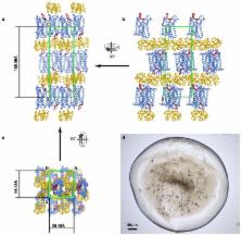
          CC chemokine receptor 2 (CCR2) is one of 19 members of the chemokine receptor subfamily of human Class A G protein-coupled receptors (GPCRs). CCR2 is expressed on monocytes, immature dendritic cells and T cell subpopulations, and mediates their migration towards endogenous CC chemokine ligands such as CCL2 1 . CCR2 and its ligands are implicated in numerous inflammatory and neurodegenerative diseases 2 including atherosclerosis, multiple sclerosis, asthma, neuropathic pain, and diabetic nephropathy, as well as cancer 3 . These disease associations have motivated numerous preclinical studies and clinical trials 4 (see ClinicalTrials.gov) in search of therapies that target the CCR2:chemokine axis. To aid drug discovery efforts 5 , we solved a structure of CCR2 in a ternary complex with an orthosteric (BMS-681 6 ) and allosteric (CCR2-RA-[ R] 7 ) antagonist. BMS-681 inhibits chemokine binding by occupying the orthosteric pocket of the receptor in a previously unseen binding mode. CCR2-RA-[ R] binds in a novel, highly druggable pocket that is the most intracellular allosteric site observed in Class A GPCRs to date; this site spatially overlaps the G protein-binding site in homologous receptors. CCR2-RA-[ R] inhibits CCR2 non-competitively by blocking activation-associated conformational changes and formation of the G protein-binding interface. The conformational signature of the conserved microswitch residues observed in double-antagonist-bound CCR2 resembles the most inactive GPCR structures solved to date. Like other protein:protein interactions, receptor:chemokine complexes are considered challenging therapeutic targets for small molecules, and the present structure suggests diverse pocket epitopes that can be exploited to overcome drug design obstacles.

          Related collections

          Most cited references32

          • Record: found
          • Abstract: found
          • Article: not found

          Crystal structure of rhodopsin: A G protein-coupled receptor.

          Heterotrimeric guanine nucleotide-binding protein (G protein)-coupled receptors (GPCRs) respond to a variety of different external stimuli and activate G proteins. GPCRs share many structural features, including a bundle of seven transmembrane alpha helices connected by six loops of varying lengths. We determined the structure of rhodopsin from diffraction data extending to 2.8 angstroms resolution. The highly organized structure in the extracellular region, including a conserved disulfide bridge, forms a basis for the arrangement of the seven-helix transmembrane motif. The ground-state chromophore, 11-cis-retinal, holds the transmembrane region of the protein in the inactive conformation. Interactions of the chromophore with a cluster of key residues determine the wavelength of the maximum absorption. Changes in these interactions among rhodopsins facilitate color discrimination. Identification of a set of residues that mediate interactions between the transmembrane helices and the cytoplasmic surface, where G-protein activation occurs, also suggests a possible structural change upon photoactivation.
            Bookmark
            • Record: found
            • Abstract: found
            • Article: not found

            Structures of the CXCR4 chemokine GPCR with small-molecule and cyclic peptide antagonists.

            Chemokine receptors are critical regulators of cell migration in the context of immune surveillance, inflammation, and development. The G protein-coupled chemokine receptor CXCR4 is specifically implicated in cancer metastasis and HIV-1 infection. Here we report five independent crystal structures of CXCR4 bound to an antagonist small molecule IT1t and a cyclic peptide CVX15 at 2.5 to 3.2 angstrom resolution. All structures reveal a consistent homodimer with an interface including helices V and VI that may be involved in regulating signaling. The location and shape of the ligand-binding sites differ from other G protein-coupled receptors and are closer to the extracellular surface. These structures provide new clues about the interactions between CXCR4 and its natural ligand CXCL12, and with the HIV-1 glycoprotein gp120.
              Bookmark
              • Record: found
              • Abstract: found
              • Article: not found

              Crystal structure of the human beta2 adrenergic G-protein-coupled receptor.

              Structural analysis of G-protein-coupled receptors (GPCRs) for hormones and neurotransmitters has been hindered by their low natural abundance, inherent structural flexibility, and instability in detergent solutions. Here we report a structure of the human beta2 adrenoceptor (beta2AR), which was crystallized in a lipid environment when bound to an inverse agonist and in complex with a Fab that binds to the third intracellular loop. Diffraction data were obtained by high-brilliance microcrystallography and the structure determined at 3.4 A/3.7 A resolution. The cytoplasmic ends of the beta2AR transmembrane segments and the connecting loops are well resolved, whereas the extracellular regions of the beta2AR are not seen. The beta2AR structure differs from rhodopsin in having weaker interactions between the cytoplasmic ends of transmembrane (TM)3 and TM6, involving the conserved E/DRY sequences. These differences may be responsible for the relatively high basal activity and structural instability of the beta2AR, and contribute to the challenges in obtaining diffraction-quality crystals of non-rhodopsin GPCRs.
                Bookmark

                Author and article information

                Journal
                0410462
                6011
                Nature
                Nature
                Nature
                0028-0836
                1476-4687
                10 November 2016
                07 December 2016
                15 December 2016
                07 June 2017
                : 540
                : 7633
                : 458-461
                Affiliations
                [1 ]Skaggs School of Pharmacy and Pharmaceutical Sciences, University of California at San Diego, La Jolla, CA 92093, USA
                [2 ]Division of Medicinal Chemistry, Leiden Academic Centre for Drug Research (LACDR), Leiden University, Leiden, The Netherlands
                [3 ]Bridge Institute, Departments of Chemistry and Physics & Astronomy, University of Southern California, Los Angeles, CA 90089, USA
                [4 ]Bristol-Myers Squibb Company, Princeton, New Jersey 08543, USA
                [5 ]Vertex Pharmaceuticals Inc. San Diego, CA 92121, USA
                [6 ]The Bridge Institute, Departments of Biological Sciences and Chemistry, University of Southern California, Los Angeles, CA 90089, USA
                Author notes
                [* ]Correspondence and requests for materials should be addressed to Tracy M. Handel ( thandel@ 123456ucsd.edu ), Irina Kufareva ( ikufareva@ 123456ucsd.edu ) or Laura H. Heitman ( l.h.heitman@ 123456lacdr.leidenuniv.nl )
                [1]

                University of California, San Diego, Skaggs School of Pharmacy and Pharmaceutical Sciences, La Jolla, CA 92093, USA.

                Yi Zheng, Ling Qin, Martin Gustavsson, Chunxia Zhao, Ruben Abagyan, Irina Kufareva, Tracy M. Handel

                [2]

                Division of Medicinal Chemistry, Leiden Academic Centre for Drug Research (LACDR), Leiden University, Leiden, The Netherlands

                Natalia V. Ortiz Zacarías, Henk de Vries, Adriaan P. IJzerman, Laura H. Heitman,

                [3]

                Bridge Institute, Departments of Chemistry, Biological Sciences and Physics & Astronomy, University of Southern California, Los Angeles, CA 90089, USA

                Gye Won Han, Vadim Cherezov

                [4]

                Bristol-Myers Squibb Company, Princeton, New Jersey 08543, USA.

                Marta Dabros, Robert J. Cherney, Percy Carter, Andrew Tebben

                [5]

                Vertex Pharmaceuticals Inc. San Diego, CA 92121, USA

                Dean Stamos

                [6]

                The Bridge Institute, Departments of Biological Sciences and Chemistry, University of Southern California, Los Angeles, CA 90089, USA

                Raymond C. Stevens

                Article
                NIHMS828647
                10.1038/nature20605
                5159191
                27926736
                7b046da5-b38d-4ac0-b00e-af5f2392324c

                Users may view, print, copy, and download text and data-mine the content in such documents, for the purposes of academic research, subject always to the full Conditions of use: http://www.nature.com/authors/editorial_policies/license.html#terms

                Reprints and permissions information is available at www.nature.com/reprints

                History
                Categories
                Article

                Uncategorized
                Uncategorized

                Comments

                Comment on this article