1
views
0
recommends
+1 Recommend
0 collections
    0
    shares
      • Record: found
      • Abstract: found
      • Article: found
      Is Open Access

      Imaging in children with ataxia-telangiectasia—The radiologist’s approach

      research-article

      Read this article at

      Bookmark
          There is no author summary for this article yet. Authors can add summaries to their articles on ScienceOpen to make them more accessible to a non-specialist audience.

          Abstract

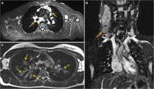
          Ataxia-telangiectasia (A-T) is a syndromic inborn error of immunity (IEI) characterized by genomic instability, defective reparation of the DNA double-strand breaks, and hypersensitivity to ionizing radiation disturbing cellular homeostasis. The role of imaging diagnostics and the conscious choice of safe and advantageous imaging technique, as well as its correct interpretation, are crucial in the diagnostic process and monitoring of children with A-T. This study aimed at defining the role of a radiologist in the early diagnosis of A-T, as well as in detecting and tracking disease complications associated with infections, inflammation, lymphoproliferation, organ-specific immunopathology, and malignancy. Based on our single-center experience, retrospective analysis of investigations using ionizing radiation-free techniques, ultrasound (US), and Magnetic Resonance Imaging (MRI), was performed on regularly followed-up 11 pediatric A-T patients, 6 girls and 5 boys, aged from 2 to 18 years, with the longest period of observation coming to over 13 years. Our attention was especially drawn to the abnormalities that were observed in the US and MRI examinations of the lungs, abdominal cavity, and lymph nodes. The abdominal US showed no abnormalities in organ dimensions or echostructure in 4 out of 11 children studied, yet in the other 7, during follow-up examinations, hepato- and/or splenomegaly, mesenteric, visceral, and paraaortic lymphadenopathy were observable. In 2 patients, focal changes in the liver and spleen were shown, and in one patient progressive abdominal lymphadenopathy corresponded with the diagnosis of non-Hodgkin lymphoma (NHL). The lung US revealed multiple subpleural consolidations and B line artifacts related to the interstitial-alveolar syndrome in 5 patients, accompanied by pleural effusion in one of them. The MRI investigation of the lung enabled the detection of lymphatic nodal masses in the mediastinum, with concomitant airway lesions characteristic of bronchiectasis and focal parenchymal consolidations in one A-T patient with chronic respiratory failure. This patient also manifested organomegaly and granulomatous liver disease in abdominal MRI examination. Our study shows that the use of modern US capabilities and MRI is safe and efficient, thereby serving as a recommended advantageous imaging diagnostic tool in monitoring children with IEI and DNA instability syndromes.

          Related collections

          Most cited references52

          • Record: found
          • Abstract: not found
          • Article: not found

          Cellular functions of the protein kinase ATM and their relevance to human disease

            Bookmark
            • Record: found
            • Abstract: found
            • Article: not found

            Antibody deficiency in patients with ataxia telangiectasia is caused by disturbed B- and T-cell homeostasis and reduced immune repertoire diversity.

            Ataxia telangiectasia (AT) is a multisystem DNA-repair disorder caused by mutations in the ataxia telangiectasia mutated (ATM) gene. Patients with AT have reduced B- and T-cell numbers and a highly variable immunodeficiency. ATM is important for V(D)J recombination and immunoglobulin class-switch recombination (CSR); however, little is known about the mechanisms resulting in antibody deficiency severity.
              Bookmark
              • Record: found
              • Abstract: found
              • Article: not found

              Lung ultrasound for the diagnosis of community-acquired pneumonia in children

              Ultrasound (US) has been proposed as an alternative first-line imaging modality to diagnose community-acquired pneumonia in children. Lung US has the potential benefits over chest radiography of being radiation free, subject to fewer regulatory requirements, relatively lower cost and with immediate bedside availability of results. However, the uptake of lung US into clinical practice has been slow and it is not yet included in clinical guidelines for community-acquired pneumonia in children. The aim of this review is to give an overview of the equipment and techniques used to perform lung US in children with suspected pneumonia and the interpretation of relevant sonographic findings. We also summarise the current evidence of diagnostic accuracy and reliability of lung US compared to alternative imaging modalities in children and critically consider the strengths and limitations of lung US for use in children presenting with suspected community-acquired pneumonia.
                Bookmark

                Author and article information

                Contributors
                URI : http://loop.frontiersin.org/people/1838496/overview
                URI : http://loop.frontiersin.org/people/1980973/overview
                URI : http://loop.frontiersin.org/people/860525/overview
                Journal
                Front Pediatr
                Front Pediatr
                Front. Pediatr.
                Frontiers in Pediatrics
                Frontiers Media S.A.
                2296-2360
                16 September 2022
                2022
                : 10
                : 988645
                Affiliations
                [1] 1Department of Pediatric Radiology, Institute of Pediatrics, Pozna University of Medical Sciences , Poznań, Poland
                [2] 2Department of Radiology, Greater Poland Cancer Center , Poznań, Poland
                [3] 3Department of Pediatric Pneumonology, Allergy and Clinical Immunology, Institute of Pediatrics, Poznań University of Medical Sciences , Poznań, Poland
                Author notes

                Edited by: Rita Consolini, University of Pisa, Italy

                Reviewed by: Martin Francis Lavin, The University of Queensland, Australia; Andreea Nissenkorn, Wolfson Medical Center, Israel; Sharon McGrath-Morrow, Children’s Hospital of Philadelphia, United States

                *Correspondence: Katarzyna Jończyk-Potoczna, jonczyk@ 123456ump.edu.pl

                This article was submitted to Pediatric Immunology, a section of the journal Frontiers in Pediatrics

                Article
                10.3389/fped.2022.988645
                9523007
                36186632
                8031c267-bb53-450e-8bd0-3297c2b3084a
                Copyright © 2022 Jończyk-Potoczna, Potoczny and Szczawińska-Popłonyk.

                This is an open-access article distributed under the terms of the Creative Commons Attribution License (CC BY). The use, distribution or reproduction in other forums is permitted, provided the original author(s) and the copyright owner(s) are credited and that the original publication in this journal is cited, in accordance with accepted academic practice. No use, distribution or reproduction is permitted which does not comply with these terms.

                History
                : 07 July 2022
                : 29 August 2022
                Page count
                Figures: 4, Tables: 4, Equations: 0, References: 52, Pages: 10, Words: 5895
                Categories
                Pediatrics
                Original Research

                imaging,ataxia-telangiectasia,magnetic resonance,ultrasound,lymphadenopathy,children

                Comments

                Comment on this article