11
views
0
recommends
+1 Recommend
0 collections
    0
    shares
      • Record: found
      • Abstract: found
      • Article: found
      Is Open Access

      Platelet-derived growth factor receptor-α and -β promote cancer stem cell phenotypes in sarcomas

      research-article

      Read this article at

      Bookmark
          There is no author summary for this article yet. Authors can add summaries to their articles on ScienceOpen to make them more accessible to a non-specialist audience.

          Abstract

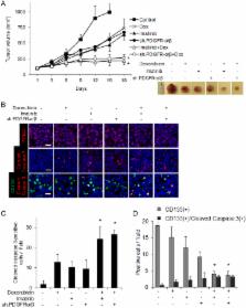
          Sarcomas are malignant tumors derived from mesenchymal tissues and may harbor a subset of cells with cancer stem-like cell (CSC) properties. Platelet-derived growth factor receptors α and β (PDGFR-α/β) play an important role in the maintenance of mesenchymal stem cells. Here we examine the role of PDGFR-α/β in sarcoma CSCs. PDGFR-α/β activity and the effects of PDGFR-α/β inhibition were examined in 3 human sarcoma cell lines using in vitro assays and mouse xenograft models. In all three cell lines, PDGFR-α/β activity was significantly higher in cells grown as spheroids (to enrich for CSCs) and in cells sorted for CD133 expression (a marker of sarcoma CSCs). Self-renewal transcription factors Nanog, Oct4, and Slug and epithelial-to-mesenchymal transition (EMT) proteins Snail, Slug, and Zeb1 were also significantly higher in spheroids cells and CD133( +) cells. Spheroid cells and CD133( +) cells demonstrated 2.9- to 4.2-fold greater migration and invasion and resistance to doxorubicin chemotherapy. Inhibition of PDGFR-α/β in CSCs using shRNA or pharmacologic inhibitors reduced expression of certain self-renewal and EMT proteins, reduced spheroid formation by 74–82%, reduced migration and invasion by 73–80%, and reversed chemotherapy resistance. In mouse xenograft models, combining PDGFR-α/β inhibition (using shRNA or imatinib) with doxorubicin had a more-than-additive effect in blocking tumor growth, with enhanced apoptosis, especially in CD133( +) cells. These results indicate that PDGFR-α/β activity is upregulated in sarcoma CSCs and promote CSC phenotypes including migration, invasion, and chemotherapy resistance. Thus, the PDGFR-α/β pathway represents a new potential therapeutic target to reduce metastatic potential and increase chemosensitivity.

          Related collections

          Most cited references19

          • Record: found
          • Abstract: found
          • Article: not found

          PDGF, TGF-beta, and FGF signaling is important for differentiation and growth of mesenchymal stem cells (MSCs): transcriptional profiling can identify markers and signaling pathways important in differentiation of MSCs into adipogenic, chondrogenic, and osteogenic lineages.

          We compared the transcriptomes of marrow-derived mesenchymal stem cells (MSCs) with differentiated adipocytes, osteocytes, and chondrocytes derived from these MSCs. Using global gene-expression profiling arrays to detect RNA transcripts, we have identified markers that are specific for MSCs and their differentiated progeny. Further, we have also identified pathways that MSCs use to differentiate into adipogenic, chondrogenic, and osteogenic lineages. We identified activin-mediated transforming growth factor (TGF)-beta signaling, platelet-derived growth factor (PDGF) signaling and fibroblast growth factor (FGF) signaling as the key pathways involved in MSC differentiation. The differentiation of MSCs into these lineages is affected when these pathways are perturbed by inhibitors of cell surface receptor function. Since growth and differentiation are tightly linked processes, we also examined the importance of these 3 pathways in MSC growth. These 3 pathways were necessary and sufficient for MSC growth. Inhibiting any of these pathways slowed MSC growth, whereas a combination of TGF-beta, PDGF, and beta-FGF was sufficient to grow MSCs in a serum-free medium up to 5 passages. Thus, this study illustrates it is possible to predict signaling pathways active in cellular differentiation and growth using microarray data and experimentally verify these predictions.
            Bookmark
            • Record: found
            • Abstract: found
            • Article: not found

            Founder and recurrent CDH1 mutations in families with hereditary diffuse gastric cancer.

            Hereditary diffuse gastric cancer is caused by germline mutations in the epithelial cadherin (CDH1) gene and is characterized by an increased risk for diffuse gastric cancer and lobular breast cancer. To determine whether recurring germline CDH1 mutations occurred due to independent mutational events or common ancestry. Thirty-eight families diagnosed clinically with hereditary diffuse gastric cancer were accrued between November 2004 and January 2006 and were analyzed for CDH1 mutations as part of an ongoing study at the British Columbia Cancer Agency. Twenty-six families had at least 2 gastric cancer cases with 1 case of diffuse gastric cancer in a person younger than 50 years; 12 families had either a single case of diffuse gastric cancer diagnosed in a person younger than 35 years or multiple cases of diffuse gastric cancer diagnosed in persons older than 50 years. Classification of family members as carriers or noncarriers of CDH1 mutations. Haplotype analysis to assess recurring mutations for common ancestry was performed on 7 families from this study and 7 previously reported families with the same mutations. Thirteen mutations (6 novel) were identified in 15 of the 38 families (40% detection rate). The 1137G>A splicing mutation and the 1901C>T (A634V) missense/splicing mutation occurred on common haplotypes in 2 families but on different haplotypes in a third family. The 2195G>A (R732Q) missense/splicing mutation occurred in 2 families on different haplotypes. The 2064-2065delTG mutation occurred on a common haplotype in 2 families. Two families from this study plus 2 additional families carrying the novel 2398delC mutation shared a common haplotype, suggesting a founder effect. All 4 families originate from the southeast coast of Newfoundland. Due to concentrations of lobular breast cancer cases, 2 branches of this family had been diagnosed as having hereditary breast cancer and were tested for BRCA mutations. Within these 4 families, the cumulative risk by age 75 years in mutation carriers for clinically detected gastric cancer was 40% (95% confidence interval [CI], 12%-91%) for males and 63% (95% CI, 19%-99%) for females and the risk for breast cancer in female mutation carriers was 52% (95% CI, 29%-94%). Recurrent CDH1 mutations in families with hereditary diffuse gastric cancer are due to both independent mutational events and common ancestry. The presence of a founder mutation from Newfoundland is strongly supported.
              Bookmark
              • Record: found
              • Abstract: found
              • Article: not found

              The cancer stem cell hypothesis: a work in progress.

              There is a growing body of evidence that supports the idea that malignant tumors are initiated and maintained by a population of tumor cells that share similar biologic properties to normal adult stem cells. This model, the cancer stem cell (CSC) hypothesis, is based on the observation that tumors, like adult tissues, arise from cells that exhibit the ability to self-renew as well as give rise to differentiated tissue cells. Although the concept of the CSC is not entirely new, advances made over the past two decades in our understanding of normal stem cell biology in conjunction with the recent application of these concepts to experimentally define CSCs have resulted in the identification of CSCs in several human malignancies.
                Bookmark

                Author and article information

                Contributors
                +212 639 7436 , yoons@mskcc.org
                Journal
                Oncogenesis
                Oncogenesis
                Oncogenesis
                Nature Publishing Group UK (London )
                2157-9024
                19 June 2018
                19 June 2018
                June 2018
                : 7
                : 6
                : 47
                Affiliations
                [1 ]ISNI 0000 0001 2171 9952, GRID grid.51462.34, Department of Surgery, , Memorial Sloan Kettering Cancer Center, ; New York, NY USA
                [2 ]ISNI 0000 0001 2171 9952, GRID grid.51462.34, Department of Medicine, , Memorial Sloan Kettering Cancer Center, ; New York, NY USA
                [3 ]ISNI 0000 0004 1936 8972, GRID grid.25879.31, Abramson Family Cancer Research Institute, Perelman School of Medicine, , University of Pennsylvania, ; Philadelphia, PA 19104 USA
                Author information
                http://orcid.org/0000-0003-4462-4301
                Article
                59
                10.1038/s41389-018-0059-1
                6006341
                29915281
                804f985c-970a-4345-9454-43a000851ecd
                © The Author(s) 2018

                Open Access This article is licensed under a Creative Commons Attribution 4.0 International License, which permits use, sharing, adaptation, distribution and reproduction in any medium or format, as long as you give appropriate credit to the original author(s) and the source, provide a link to the Creative Commons license, and indicate if changes were made. The images or other third party material in this article are included in the article’s Creative Commons license, unless indicated otherwise in a credit line to the material. If material is not included in the article’s Creative Commons license and your intended use is not permitted by statutory regulation or exceeds the permitted use, you will need to obtain permission directly from the copyright holder. To view a copy of this license, visit http://creativecommons.org/licenses/by/4.0/.

                History
                : 2 May 2018
                : 21 May 2018
                : 21 May 2018
                Categories
                Article
                Custom metadata
                © The Author(s) 2018

                Oncology & Radiotherapy
                Oncology & Radiotherapy

                Comments

                Comment on this article