13
views
0
recommends
+1 Recommend
0 collections
    0
    shares
      • Record: found
      • Abstract: found
      • Article: found
      Is Open Access

      The Actin Cytoskeleton Is Involved in Glial Cell Line-Derived Neurotrophic Factor (GDNF)-Induced Ret Translocation into Lipid Rafts in Dopaminergic Neuronal Cells

      research-article

      Read this article at

      Bookmark
          There is no author summary for this article yet. Authors can add summaries to their articles on ScienceOpen to make them more accessible to a non-specialist audience.

          Abstract

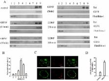
          Glial cell line-derived neurotrophic factor (GDNF), a potential therapeutic factor for Parkinson’s disease (PD), exerts its biological effects through the Ret receptor tyrosine kinase. The redistribution of Ret into lipid rafts substantially influences Ret signaling, but the mechanisms underlying Ret translocation remain unclear. The purpose of our study was to further explore the signaling mechanisms of GDNF and to determine whether the actin cytoskeleton is involved in the GDNF-induced Ret translocation into lipid rafts. In MN9D dopaminergic neuronal cells, we used density gradient centrifugation and immunofluorescence confocal microscopy to separate and visualize lipid rafts, co-immunoprecipitation to analyze protein-protein interactions, and latrunculin B (Lat B) and jasplakinolide (Jas) to disrupt and enhance the polymerization of the actin cytoskeleton, respectively. The results showed that Ret translocated into lipid rafts and coimmunoprecipitated with actin in response to GDNF treatment. After Lat B or Jas treatment, the Ret–F-actin association induced by GDNF was impaired or enhanced respectively and then the levels of Ret translocated into lipid rafts were correspondingly inhibited or promoted. These data indicate that actin polymerization and cytoskeletal remodeling are integral to GDNF-induced cell signaling in dopaminergic cells and define a new role of the actin cytoskeleton in promoting Ret redistribution into lipid rafts.

          Related collections

          Most cited references18

          • Record: found
          • Abstract: found
          • Article: not found

          Active remodeling of cortical actin regulates spatiotemporal organization of cell surface molecules.

          Many lipid-tethered proteins and glycolipids exist as monomers and nanoclusters on the surface of living cells. The spatial distribution and dynamics of formation and breakup of nanoclusters does not reflect thermal and chemical equilibrium and is controlled by active remodeling of the underlying cortical actin. We propose a model for nanoclustering based on active hydrodynamics, wherein cell surface molecules bound to dynamic actin are actively driven to form transient clusters. This consistently explains all of our experimental observations. Using FCS and TIRF microscopy, we provide evidence for the existence of short, dynamic, polymerizing actin filaments at the cortex, a key assumption of the theoretical framework. Our theory predicts that lipid-anchored proteins that interact with dynamic actin must exhibit anomalous concentration fluctuations, and a cell membrane protein capable of binding directly to actin can form nanoclusters. These we confirm experimentally, providing an active mechanism for molecular organization and its spatiotemporal regulation on the plasma membrane. Copyright © 2012 Elsevier Inc. All rights reserved.
            Bookmark
            • Record: found
            • Abstract: found
            • Article: not found

            Aggregation of Lipid Rafts Accompanies Signaling via the T Cell Antigen Receptor

            The role of lipid rafts in T cell antigen receptor (TCR) signaling was investigated using fluorescence microscopy. Lipid rafts labeled with cholera toxin B subunit (CT-B) and cross-linked into patches displayed characteristics of rafts isolated biochemically, including detergent resistance and colocalization with raft-associated proteins. LCK, LAT, and the TCR all colocalized with lipid patches, although TCR association was sensitive to nonionic detergent. Aggregation of the TCR by anti-CD3 mAb cross-linking also caused coaggregation of raft-associated proteins. However, the protein tyrosine phosphatase CD45 did not colocalize to either CT-B or CD3 patches. Cross-linking of either CD3 or CT-B strongly induced tyrosine phosphorylation and recruitment of a ZAP-70(SH2)2–green fluorescent protein (GFP) fusion protein to the lipid patches. Also, CT-B patching induced signaling events analagous to TCR stimulation, with the same dependence on expression of key TCR signaling molecules. Targeting of LCK to rafts was necessary for these events, as a nonraft- associated transmembrane LCK chimera, which did not colocalize with TCR patches, could not reconstitute CT-B–induced signaling. Thus, our results indicate a mechanism whereby TCR engagement promotes aggregation of lipid rafts, which facilitates colocalization of LCK, LAT, and the TCR whilst excluding CD45, thereby triggering protein tyrosine phosphorylation.
              Bookmark
              • Record: found
              • Abstract: found
              • Article: not found

              Dynamic molecular confinement in the plasma membrane by microdomains and the cytoskeleton meshwork.

              It is by now widely recognized that cell membranes show complex patterns of lateral organization. Two mechanisms involving either a lipid-dependent (microdomain model) or cytoskeleton-based (meshwork model) process are thought to be responsible for these plasma membrane organizations. In the present study, fluorescence correlation spectroscopy measurements on various spatial scales were performed in order to directly identify and characterize these two processes in live cells with a high temporal resolution, without any loss of spatial information. Putative raft markers were found to be dynamically compartmented within tens of milliseconds into small microdomains (Ø <120 nm) that are sensitive to the cholesterol and sphingomyelin levels, whereas actin-based cytoskeleton barriers are responsible for the confinement of the transferrin receptor protein. A free-like diffusion was observed when both the lipid-dependent and cytoskeleton-based organizations were disrupted, which suggests that these are two main compartmentalizing forces at work in the plasma membrane.
                Bookmark

                Author and article information

                Journal
                Int J Mol Sci
                Int J Mol Sci
                ijms
                International Journal of Molecular Sciences
                MDPI
                1422-0067
                07 September 2017
                September 2017
                : 18
                : 9
                : 1922
                Affiliations
                [1 ]Department of Pathophysiology, School of the Basic Medicine, Xuzhou Medical University, Xuzhou 221004, Jiangsu, China; xzmclili@ 123456163.com (L.L.); xzmcxm@ 123456163.com (M.X.); xzmclcx@ 123456163.com (C.L.); xzmcwy@ 123456163.com (Y.W.); xzmcqys@ 123456163.com (Y.Q.)
                [2 ]Institute of Emergency Rescue Medicine, Xuzhou Medical University, Xuzhou 221004, Jiangsu, China; xzmcshj@ 123456163.com
                [3 ]School of Nursing, Xuzhou Medical University, Xuzhou 221004, Jiangsu, China; xzyxympp@ 123456163.com
                [4 ]Department of Histology and Embryology, School of the Basic Medicine, Xuzhou Medical University, Xuzhou 221004, Jiangsu, China; 5189009@ 123456xzhmu.edu.cn
                [5 ]Department of Neurobiology and Anatomy, Xuzhou Key Laboratory of Neurobiology, Jiangsu Key Laboratory of New Drug Research and Clinical Pharmacy, Xuzhou Medical University, Xuzhou 221004, Jiangsu, China; xzmcgj@ 123456163.com
                [6 ]Institute of Medical Technology, Xuzhou Medical University, Xuzhou 221004, Jiangsu, China; wt27518@ 123456sina.com
                Author notes
                [* ]Correspondence: gds@ 123456xzhmu.edu.cn ; Tel.: +86-516-8326-2301
                Author information
                https://orcid.org/0000-0001-8567-0238
                Article
                ijms-18-01922
                10.3390/ijms18091922
                5618571
                28880247
                8130788e-89fb-49d0-924a-8d552071fcbc
                © 2017 by the authors.

                Licensee MDPI, Basel, Switzerland. This article is an open access article distributed under the terms and conditions of the Creative Commons Attribution (CC BY) license ( http://creativecommons.org/licenses/by/4.0/).

                History
                : 06 July 2017
                : 05 September 2017
                Categories
                Article

                Molecular biology
                gdnf,ret,dopaminergic cells,lipid rafts,cytoskeleton
                Molecular biology
                gdnf, ret, dopaminergic cells, lipid rafts, cytoskeleton

                Comments

                Comment on this article