2
views
0
recommends
+1 Recommend
0 collections
    0
    shares
      • Record: found
      • Abstract: found
      • Article: found
      Is Open Access

      Disseminated Juvenile Xanthogranuloma and Hemophagocytic Lymphohistiocytosis Developed During Treatment of Acute Lymphoblastic Leukemia: Case Report

      case-report

      Read this article at

      Bookmark
          There is no author summary for this article yet. Authors can add summaries to their articles on ScienceOpen to make them more accessible to a non-specialist audience.

          Abstract

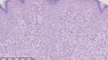
          The association between acute lymphoblastic leukemia (ALL), non-Langerhans cell histiocytosis (non-LCH), and hemophagocytic lymphohistiocytosis (HLH), to the best of our knowledge, has not been published to date. Juvenile xanthogranuloma (JXG), as a type of non-LCH, is usually a benign disease limited to the skin. Systemic involvement is rarely reported. The present case report describes a 15-year-old boy diagnosed with disseminated JXG involving skin and bone marrow concurrent with severe symptoms of HLH during ALL therapy. Examination of immunoglobulin heavy chain genes in B-cell precursor leukemic blasts and histiocytes in the skin and bone marrow revealed identical rearrangements, confirming clonal relationship between both diseases. Implementation of corticosteroids, vinblastine, etoposide, cyclosporine, and tocilizumab resulted in partial skin lesion resolution with no improvement of bone marrow function; therefore, hematopoietic stem cell transplantation (HSCT) was eventually performed. The patient's hematological and general status has improved gradually; however, remarkable recovery of skin lesions was observed after empirical antitubercular therapy. Mycobacterium spp. infection should be considered as a possible secondary HLH trigger. Triple association of ALL, non-LCH, and HLH highlights heterogeneity of histiocytic disorders and possible common origin of dendritic and lymphoid cells.

          Related collections

          Most cited references14

          • Record: found
          • Abstract: found
          • Article: not found

          Severe imbalance of IL-18/IL-18BP in patients with secondary hemophagocytic syndrome.

          Hemophagocytic syndrome (HPS) is characterized by an uncontrolled and poorly understood activation of T-helper 1 (Th-1) lymphocytes and macrophages. We studied 20 patients with HPS secondary to infections, autoimmune disease, lymphoma, or cancer and observed that the concentrations of serum interleukin 18 (IL-18), a strong inducer of Th-1 responses, interferon gamma (IFN-gamma) production, and stimulation of macrophages and natural killer (NK) cells were highly increased in HPS but not in control patients. In contrast, concentrations of its natural inhibitor, the IL-18 binding protein (IL-18BP), were only moderately elevated, resulting in a high level of biologically active free IL-18 in HPS (4.6-fold increase compared with controls; P < .001). Free IL-18 but not IL-12 concentrations significantly correlated with clinical status and the biologic markers of HPS such as anemia (P < .001), hypertriglyceridemia, and hyperferritinemia (P < .01) and also with markers of Th-1 lymphocyte or macrophage activation, such as elevated concentrations of IFN-gamma and soluble IL-2 and tumor necrosis factor alpha (TNF-alpha) receptor concentrations. Despite high IL-18 elevation, in vitro NK-cell cytotoxicity was severely impaired in HPS patients, in part due to NK-cell lymphopenia that was observed in a majority of patients but also secondary to an intrinsic NK-cell functional deficiency. We concluded that a severe IL-18/IL-18BP imbalance results in Th-1 lymphocyte and macrophage activation, which escapes control by NK-cell cytotoxicity and may allow for secondary HPS in patients with underlying diseases.
            Bookmark
            • Record: found
            • Abstract: found
            • Article: not found

            Understanding organ dysfunction in hemophagocytic lymphohistiocytosis.

            This review aims to help critical care clinicians maintain a high level of suspicion regarding the diagnosis of Hemophagocytic Histiolymphocytosis (HLH). It describes the clinical and laboratory features of HLH, outlines its pathophysiology and reviews the most frequent etiologies related to HLH. Prognostic factors and therapeutic options are also reported. Review of the literature. The diagnosis of HLH relies on the association of clinical abnormalities and hemophagocytosis in bone marrow, spleen, or lymph node specimens. Liver, pulmonary, renal, cardiac and skin involvement may occur at various degrees possibly leading to multiple organ failure. Three main etiologies can be found, namely infections, lymphoproliferative diseases, or connective tissue diseases. Immune deficiency is often retrieved. Mortality can be as high as 50%. Although clinically mimicking severe sepsis, HLH has a distinct pathophysiology on which specific therapy is based. Early diagnosis and treatment is mandatory to increase the chances of survival. The comprehensive management of severe HLH requires the involvement of a multidisciplinary team in order to determine the best therapeutic strategy and to identify the underlying cause.
              Bookmark
              • Record: found
              • Abstract: found
              • Article: found
              Is Open Access

              Malignancy and chemotherapy induced haemophagocytic lymphohistiocytosis in children and adolescents—a single centre experience of 20 years

              Haemophagocytic lymphohistiocytosis (HLH) is a possibly life-threatening syndrome of immune dysregulation and can be divided into primary (hereditary) and secondary forms (including malignancy-associated HLH (M-HLH)). We retrospectively analysed epidemiological, clinical, virological and laboratory data from patients with M-HLH treated at our department between 1995 and 2014. Out of 1.706 haemato-/oncologic patients treated at our department between 1995 and 2014, we identified 22 (1.29%) patients with secondary HLH (1.3–18.0, median 10.1 years; malignancy induced n = 2; chemotherapy induced n = 20). Patients with acute myeloblastic leukaemia (AML) developed HLH significantly more often than patients with acute lymphoblastic leukaemia (ALL) (10/55, 18.2% vs. 6/148, 4.1%, p = 0.0021). As possible viral triggers, we detected BKV (53.8% of the tested patients), HHV-6 (33.3%), EBV (27.8%), CMV (23.5%), ADV (16.7%) and PVB19 (16.7%) significantly more frequently than in haemato-/oncologic patients without HLH. Despite lacking evidence of concurrent bacterial infection, C-reactive protein (CRP) and procalcitotnin (PCT) were elevated in 94.7 and 77.7% of the patients, respectively. Ferritin and sIL2R were markedly elevated in all patients. HLH-associated mortality significantly (p = 0.0276) decreased from 66.6% (1995–2004) to 6.25% (2005–2014), suggesting improved diagnostic and therapeutic management. Awareness of HLH is important, and fever refractory to antibiotics should prompt to consider this diagnosis. Elevated ferritin and sIL2R seem to be good markers, while inflammatory markers like CRP and PCT are not useful to discriminate viral triggered HLH from severe bacterial infection. Re-/activation of several viruses may play a role as possible trigger.
                Bookmark

                Author and article information

                Contributors
                Journal
                Front Oncol
                Front Oncol
                Front. Oncol.
                Frontiers in Oncology
                Frontiers Media S.A.
                2234-943X
                03 July 2020
                2020
                : 10
                : 921
                Affiliations
                [1] 1Department of Pediatric Oncology and Hematology, Faculty of Medicine, Jagiellonian University Medical College , Krakow, Poland
                [2] 2Department of Oncology and Hematology, University Children's Hospital of Krakow , Krakow, Poland
                [3] 3Department of Clinical Immunology and Transplantation, Institute of Pediatrics, Jagiellonian University Medical College , Krakow, Poland
                [4] 4Department of Clinical Immunology and Transplantology, Stem Cell Transplant Center, University Children's Hospital of Krakow, Jagiellonian University Medical College , Krakow, Poland
                [5] 5Department of Pathology, University Children's Hospital of Krakow , Krakow, Poland
                [6] 6Department of Biochemistry, University Children's Hospital of Krakow , Krakow, Poland
                [7] 7Student Scientific Group of Pediatric Oncology and Hematology, Jagiellonian University Medical College , Krakow, Poland
                Author notes

                Edited by: Shimin Hu, University of Texas MD Anderson Cancer Center, United States

                Reviewed by: Habibe Kurt, Brown University, United States; Mina Luqing Xu, Yale University, United States

                *Correspondence: Szymon Skoczen szymon.skoczen@ 123456uj.edu.pl

                This article was submitted to Hematologic Malignancies, a section of the journal Frontiers in Oncology

                Article
                10.3389/fonc.2020.00921
                7350519
                81866c4a-857d-407c-a1ae-e866fac2bfe2
                Copyright © 2020 Pawińska-Wa̧sikowska, Cwiklinska, Wyrobek, Balwierz, Bukowska-Strakova, Dluzniewska, Gozdzik, Drabik, Rygielska, Stepien and Skoczen.

                This is an open-access article distributed under the terms of the Creative Commons Attribution License (CC BY). The use, distribution or reproduction in other forums is permitted, provided the original author(s) and the copyright owner(s) are credited and that the original publication in this journal is cited, in accordance with accepted academic practice. No use, distribution or reproduction is permitted which does not comply with these terms.

                History
                : 14 February 2020
                : 11 May 2020
                Page count
                Figures: 4, Tables: 0, Equations: 0, References: 21, Pages: 6, Words: 3924
                Categories
                Oncology
                Case Report

                Oncology & Radiotherapy
                acute lymphoblastic leukemia,non-langerhans cell histiocytosis,hemophagocytic lymphohistiocytosis,juvenile xanthogranuloma,case report

                Comments

                Comment on this article