33
views
0
recommends
+1 Recommend
0 collections
    0
    shares
      • Record: found
      • Abstract: found
      • Article: found
      Is Open Access

      Vertebral Osteomyelitis Caused by Mycobacterium abscessus Surgically Treated Using Antibacterial Iodine-Supported Instrumentation

      case-report

      Read this article at

      Bookmark
          There is no author summary for this article yet. Authors can add summaries to their articles on ScienceOpen to make them more accessible to a non-specialist audience.

          Abstract

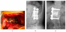
          Mycobacterium abscessus infections rarely develop in healthy individuals, and mostly they occur in immunocompromised hosts. Vertebral osteomyelitis due to Mycobacterium abscessus is very rare and only three previous cases of spinal infection caused by Mycobacterium abscessus have been reported. Mycobacterium abscessus isolates are uniformly resistant to antituberculous agents and can display a virulent biofilm-forming phenotype. The patient was a 67-year-old woman with vertebral osteomyelitis of the L1-2. She was healthy without immune-suppressed condition, history of trauma, or intravenous drug use. The smear examination of the specimen harvested by CT-guided puncture of the paravertebral abscess revealed Mycobacterium abscessus. Her disease condition did not abate with conservative treatment using antimicrobial chemotherapy. Radical debridement of the vertebral osteomyelitis and anterior reconstruction from T12 to L2 using antibacterial iodine-supported instrumentation were performed. Chemotherapy using clarithromycin, amikacin, and imipenem was applied for 6 months after surgery as these antibiotics had been proven to be effective to Mycobacterium abscessus after surgery. Two years after surgery, the infected anterior site healed and bony fusion was successfully achieved without a recurrence of infection.

          Related collections

          Most cited references14

          • Record: found
          • Abstract: found
          • Article: not found

          Molecular epidemiology of Mycobacterium abscessus, with focus on cystic fibrosis.

          Mycobacterium abscessus has been isolated increasingly often from the respiratory tracts of cystic fibrosis (CF) patients. It is not known whether these organisms are transmitted from person to person or acquired from environmental sources. Here, colony morphology and pulsed-field gel electrophoresis (PFGE) pattern were examined for 71 isolates of M. abscessus derived from 14 CF patients, three non-CF patients with chronic respiratory M. abscessus infection or colonization, one patient with mastoiditis, and four patients with infected wounds, as well as for six isolates identified as environmental contaminants in various clinical specimens. Contaminants and wound isolates mainly exhibited smooth colony morphology, while a rough colony phenotype was significantly associated with chronic airway colonization (P=0.014). Rough strains may exhibit increased airway-colonizing capacity, the cause of which remains to be determined. Examination by PFGE of consecutive isolates from the same patient showed that they all represented a single strain, even in cases where both smooth and rough isolates were present. When PFGE patterns were compared, it was shown that 24 patients had unique strains, while four patients harbored strains indistinguishable by PFGE. Two of these were siblings with CF. The other two patients, one of whom had CF, had not had contact with each other or with the siblings. Our results show that most patients colonized by M. abscessus in the airways have unique strains, indicating that these strains derive from the environment and that patient-to-patient transmission rarely occurs.
            Bookmark
            • Record: found
            • Abstract: found
            • Article: not found

            Mycobacterium abscessus: an emerging rapid-growing potential pathogen.

            Mycobacterium abscessus is the most pathogenic and chemotherapy-resistant rapid-growing mycobacterium. It is commonly associated with contaminated traumatic skin wounds and with post-surgical soft tissue infections. It is also one of the mycobacteria that are most often isolated from cystic fibrosis patients. It is essential to differentiate this species from the formerly indistinct "M. chelonae-complex", as chemotherapy is especially difficult in M. abscessussenso strictu. Clarithromycin or azithromycin are the only regular oral antimycobacterial agents with an effect on M. abscessus, and should preferably be supplemented with other drugs since long-term monotherapy may cause resistance. Amikacin is a major parenteral drug against M. abscessus that should also be given in combination with another drug. The recently introduced drug tigecycline may prove to be an important addition to chemotherapy, but has yet to be fully clinically evaluated as an antimycobacterial agent. Surgery can be curative, or at least helpful, in the healing of M. abscessus infection, and if conducted, it should include the removal of all foreign or necrotic material. There is increasing awareness of M. abscessus as an emerging pathogen.
              Bookmark
              • Record: found
              • Abstract: found
              • Article: not found

              Innovative antimicrobial coating of titanium implants with iodine

              Background Postoperative infection associated with implants remains a serious complication in orthopedic surgery. Several biomaterial surface treatments have been proposed as a means of reducing the incidence of implant-associated infections. In this study, a clinical trial was performed using an iodine-supported titanium that suppresses the microbial activities. Methods A total of 222 patients with postoperative infection or compromised status were treated using iodine-supported titanium implants. The mean age of the patients was 49.4 years (range 5–85 years). One hundred twenty-seven patients were male and 95 were female. In 158 patients, iodine-supported implants were used to prevent infection, such as compromised hosts and conditions, and in 64 patients to treat active infection. White blood cell (WBCs) and C-reactive protein (CRP) levels were measured pre- and postoperatively in all patients. To confirm whether iodine from the implant affected physiological functions, thyroid hormone levels in the blood were examined. Both examinations were conducted sequentially for a year. Radiological evaluations were performed regularly after the operation. The chronological changes of the iodine amount were evaluated using half pins removed after completion of external fixation. Results The mean follow-up period was 18.4 months (range 3–44 months). Acute infection developed in three tumor cases among the 158 patients on preventive therapy. All three recovered without removal of the implants. Infection was cured in all 64 patients with infection. Median WBC levels were in the normal range, and median CRP levels returned to <0.5 within 4 weeks after surgery. Abnormalities of thyroid gland function were not detected. None of the patients experienced loosening of the implants. There were two patients with mechanical implant failure, which was treated by re-implantation. Excellent bone ingrowth and ongrowth were found around all hip and tumor prostheses. One year later, the amount of iodine on external fixation pins remained about 20–30 %. Conclusions Iodine-supported titanium implants can be very effective for preventing and treating infections after orthopedic surgery. Cytotoxicity and adverse effects were not detected.
                Bookmark

                Author and article information

                Journal
                Case Rep Orthop
                Case Rep Orthop
                CRIOR
                Case Reports in Orthopedics
                Hindawi Publishing Corporation
                2090-6749
                2090-6757
                2014
                2 December 2014
                : 2014
                : 197061
                Affiliations
                Department of Orthopaedic Surgery, Kanazawa University School of Medicine, 13-1 Takara-machi, Kanazawa 920-8641, Japan
                Author notes

                Academic Editor: John Nyland

                Author information
                http://orcid.org/0000-0003-4762-5932
                http://orcid.org/0000-0003-0515-540X
                http://orcid.org/0000-0002-7488-3139
                http://orcid.org/0000-0003-0568-5419
                http://orcid.org/0000-0002-3415-5823
                http://orcid.org/0000-0001-5016-4917
                http://orcid.org/0000-0003-0730-7921
                Article
                10.1155/2014/197061
                4269211
                83ec6caf-e308-4a67-a5bd-659d55072e6c
                Copyright © 2014 Satoshi Kato et al.

                This is an open access article distributed under the Creative Commons Attribution License, which permits unrestricted use, distribution, and reproduction in any medium, provided the original work is properly cited.

                History
                : 18 February 2014
                : 6 November 2014
                : 13 November 2014
                Categories
                Case Report

                Orthopedics
                Orthopedics

                Comments

                Comment on this article