0
views
0
recommends
+1 Recommend
0 collections
    0
    shares
      • Record: found
      • Abstract: found
      • Article: found
      Is Open Access

      ELAVL2 loss promotes aggressive mesenchymal transition in glioblastoma

      research-article

      Read this article at

      Bookmark
          There is no author summary for this article yet. Authors can add summaries to their articles on ScienceOpen to make them more accessible to a non-specialist audience.

          Abstract

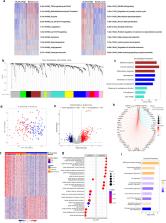
          Glioblastoma (GBM), the most lethal primary brain cancer, exhibits intratumoral heterogeneity and molecular plasticity, posing challenges for effective treatment. Despite this, the regulatory mechanisms underlying such plasticity, particularly mesenchymal (MES) transition, remain poorly understood. In this study, we elucidate the role of the RNA-binding protein ELAVL2 in regulating aggressive MES transformation in GBM. We found that ELAVL2 is most frequently deleted in GBM compared to other cancers and associated with distinct clinical and molecular features. Transcriptomic analysis revealed that ELAVL2-mediated alterations correspond to specific GBM subtype signatures. Notably, ELAVL2 expression negatively correlated with epithelial-to-mesenchymal transition (EMT)-related genes, and its loss promoted MES process and chemo-resistance in GBM cells, whereas ELAVL2 overexpression exerted the opposite effect. Further investigation via tissue microarray analysis demonstrated that high ELAVL2 protein expression confers a favorable survival outcome in GBM patients. Mechanistically, ELAVL2 was shown to directly bind to the transcripts of EMT-inhibitory molecules, SH3GL3 and DNM3, modulating their mRNA stability, potentially through an m6A-dependent mechanism. In summary, our findings identify ELAVL2 as a critical tumor suppressor and mRNA stabilizer that regulates MES transition in GBM, underscoring its role in transcriptomic plasticity and glioma progression.

          Related collections

          Most cited references59

          • Record: found
          • Abstract: found
          • Article: not found

          Gene set enrichment analysis: A knowledge-based approach for interpreting genome-wide expression profiles

          Although genomewide RNA expression analysis has become a routine tool in biomedical research, extracting biological insight from such information remains a major challenge. Here, we describe a powerful analytical method called Gene Set Enrichment Analysis (GSEA) for interpreting gene expression data. The method derives its power by focusing on gene sets, that is, groups of genes that share common biological function, chromosomal location, or regulation. We demonstrate how GSEA yields insights into several cancer-related data sets, including leukemia and lung cancer. Notably, where single-gene analysis finds little similarity between two independent studies of patient survival in lung cancer, GSEA reveals many biological pathways in common. The GSEA method is embodied in a freely available software package, together with an initial database of 1,325 biologically defined gene sets.
            Bookmark
            • Record: found
            • Abstract: found
            • Article: found
            Is Open Access

            limma powers differential expression analyses for RNA-sequencing and microarray studies

            limma is an R/Bioconductor software package that provides an integrated solution for analysing data from gene expression experiments. It contains rich features for handling complex experimental designs and for information borrowing to overcome the problem of small sample sizes. Over the past decade, limma has been a popular choice for gene discovery through differential expression analyses of microarray and high-throughput PCR data. The package contains particularly strong facilities for reading, normalizing and exploring such data. Recently, the capabilities of limma have been significantly expanded in two important directions. First, the package can now perform both differential expression and differential splicing analyses of RNA sequencing (RNA-seq) data. All the downstream analysis tools previously restricted to microarray data are now available for RNA-seq as well. These capabilities allow users to analyse both RNA-seq and microarray data with very similar pipelines. Second, the package is now able to go past the traditional gene-wise expression analyses in a variety of ways, analysing expression profiles in terms of co-regulated sets of genes or in terms of higher-order expression signatures. This provides enhanced possibilities for biological interpretation of gene expression differences. This article reviews the philosophy and design of the limma package, summarizing both new and historical features, with an emphasis on recent enhancements and features that have not been previously described.
              Bookmark
              • Record: found
              • Abstract: found
              • Article: not found

              NIH Image to ImageJ: 25 years of image analysis

              For the past twenty five years the NIH family of imaging software, NIH Image and ImageJ have been pioneers as open tools for scientific image analysis. We discuss the origins, challenges and solutions of these two programs, and how their history can serve to advise and inform other software projects.
                Bookmark

                Author and article information

                Contributors
                paeksh@snu.ac.kr
                Journal
                NPJ Precis Oncol
                NPJ Precis Oncol
                NPJ Precision Oncology
                Nature Publishing Group UK (London )
                2397-768X
                28 March 2024
                28 March 2024
                2024
                : 8
                : 79
                Affiliations
                [1 ]Department of Neurosurgery, Cancer Research Institute and Ischemic/Hypoxic Disease Institute, Seoul National University College of Medicine, ( https://ror.org/04h9pn542) Seoul, Korea
                [2 ]Interdisciplinary Program in Neuroscience, Seoul National University College of Biological Sciences, ( https://ror.org/04h9pn542) Seoul, Korea
                [3 ]Interdisciplinary Program in Caner Biology, Seoul National University College of Medicine, ( https://ror.org/04h9pn542) Seoul, Korea
                [4 ]GRID grid.412678.e, ISNI 0000 0004 0634 1623, Department of Neurosurgery, , Soonchunhyang University Seoul Hospital, ; Seoul, Korea
                [5 ]GRID grid.17635.36, ISNI 0000000419368657, Department of Radiation Oncology, , University of Minnesota Medical School, ; Minneapolis, MN 55455 USA
                [6 ]Korean Cell Line Bank, Laboratory of Cell Biology, Cancer Research Institute, Seoul National University College of Medicine, ( https://ror.org/04h9pn542) Seoul, Korea
                [7 ]Department of Pathology, Seoul National University Hospital, ( https://ror.org/01z4nnt86) Seoul, Korea
                [8 ]GRID grid.31501.36, ISNI 0000 0004 0470 5905, Advanced Institute of Convergence Technology, , Seoul National University, ; Suwon, Korea
                Author information
                http://orcid.org/0000-0002-7090-537X
                http://orcid.org/0000-0002-8681-1597
                http://orcid.org/0000-0003-3007-8653
                Article
                566
                10.1038/s41698-024-00566-1
                10978835
                38548861
                85d68848-f414-45c1-bc6e-5e4f74a572ed
                © The Author(s) 2024

                Open Access This article is licensed under a Creative Commons Attribution 4.0 International License, which permits use, sharing, adaptation, distribution and reproduction in any medium or format, as long as you give appropriate credit to the original author(s) and the source, provide a link to the Creative Commons licence, and indicate if changes were made. The images or other third party material in this article are included in the article’s Creative Commons licence, unless indicated otherwise in a credit line to the material. If material is not included in the article’s Creative Commons licence and your intended use is not permitted by statutory regulation or exceeds the permitted use, you will need to obtain permission directly from the copyright holder. To view a copy of this licence, visit http://creativecommons.org/licenses/by/4.0/.

                History
                : 16 May 2023
                : 8 March 2024
                Funding
                Funded by: FundRef https://doi.org/10.13039/501100003725, National Research Foundation of Korea (NRF);
                Award ID: 2015M3C7A1028926 & 2020M3A9G8022029
                Award Recipient :
                Funded by: FundRef https://doi.org/10.13039/501100003715, Korea Research Institute of Bioscience and Biotechnology (KRIBB);
                Award ID: KGM456212109816
                Award Recipient :
                Funded by: FundRef https://doi.org/10.13039/501100003696, Electronics and Telecommunications Research Institute (ETRI);
                Award ID: 21YB1500
                Award Recipient :
                Categories
                Article
                Custom metadata
                © Springer Nature Limited 2024

                cns cancer,prognostic markers
                cns cancer, prognostic markers

                Comments

                Comment on this article