9
views
0
recommends
+1 Recommend
0 collections
    0
    shares
      • Record: found
      • Abstract: found
      • Article: found
      Is Open Access

      Value of CT-MRI fusion in iodine-125 brachytherapy for high-grade glioma

      research-article

      Read this article at

      Bookmark
          There is no author summary for this article yet. Authors can add summaries to their articles on ScienceOpen to make them more accessible to a non-specialist audience.

          Abstract

          Purposes

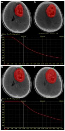
          To develop a fast, accurate and robust method of fusing Computed Tomography (CT) with pre-operative Magnetic Resonance Imaging (MRI) and evaluate the impact of using the fused data on the implantation of Iodine-125 ( 125I) seeds for brachytherapy of high-grade gliomas (HGG).

          Methods

          A study was performed on a cohort of 10 consecutive patients with HGG were treated by 125I brachytherapy with CT-MRI fusion image guided (CMGB), and 10 patients treated with CT alone guided (CGB). Statistical analysis was performed to compare (1) the planning target volume, (2) the accuracy of location of catheters, (3) the target volume covered by 150% prescribe dose ( V150), (4) the target volume covered by 200% prescribe dose ( V200), and (5) the conformity index ( CI) with or without fused data.

          Results

          The median planning target volume was 50.1 cm 3 in CGB, and 56.25 cm 3 in CMGB with significant difference ( p = 0.005). The accuracy of catheter insertion was 94.4% with CMGB and 78.9% with CGB. The median V150 and V200 was 45.32% vs 64.24% and 32.81% vs 53.17% in CGB and CMGB, respectively. There was significant difference for CI (83.5% vs. 74.5%, p < 0.05) in the two groups for the post-operative verification.

          Conclusions

          The proposed MRI-CT fusion method enables a quantitative assessment of impact on HGG brachytherapy. The additional information obtained from the fused images can be utilized for more accurate delineation of lesion boundaries and targeting of catheters. Experimental results show that the fusion algorithm is robust and reliable in clinical practice.

          Related collections

          Most cited references37

          • Record: found
          • Abstract: found
          • Article: not found

          A simple scoring ratio to index the conformity of radiosurgical treatment plans. Technical note.

          I Paddick (2000)
          A conformity index is a measure of how well the volume of a radiosurgical dose distribution conforms to the size and shape of a target volume. Because the success of radiosurgery is related to the extremely conformal irradiation of the target, an accurate method for describing this parameter is important. Existing conformity ratios and indices used in radiosurgery are reviewed and criticized. It will be demonstrated that previously proposed measurements of conformity can, under certain conditions, give false perfect scores. A new conformity index is derived that gives an objective score of conformity for a treatment plan and gives no false scores. An analysis of five different treatment plans is made using both the existing scoring methods and the new conformity index.
            Bookmark
            • Record: found
            • Abstract: not found
            • Article: not found

            Medical image fusion: A survey of the state of the art

              Bookmark
              • Record: found
              • Abstract: found
              • Article: not found

              Reirradiation of recurrent high-grade gliomas using amino acid PET (SPECT)/CT/MRI image fusion to determine gross tumor volume for stereotactic fractionated radiotherapy.

              To develop a valid treatment strategy for recurrent high-grade gliomas using stereotactic hypofractionated reirradiation based on biologic imaging and temozolomide. The trial included a total of 44 patients with recurrent high-grade gliomas (1 patient with anaplastic oligodendroglioma, 8 with anaplastic astrocytoma, 33 with glioblastoma multiforme, and 2 with gliosarcoma) after previous surgery and postoperative conventional radiotherapy +/- chemotherapy. For fractionated stereotactic radiotherapy (SFRT) treatment planning, the gross tumor volume was defined by (11)C-methionine positron emission tomography (MET-PET) or (123)I-alpha-methyl-tyrosine (IMT) single-photon computed emission tomography (SPECT)/computed tomography (CT)/magnetic resonance imaging (MRI) fusion in 82% of the patients and by CT/T1+gadolinium-MRI image fusion in 18% of the patients. Six fractions of 5 Gy were administered in 6 days. In 29 of 44 patients (66%), chemotherapy with temozolomide (200 mg/m(2) body surface/day) was given in one to two cycles before and four to five cycles after SFRT. The patients were evaluated in follow-up by clinical investigators and MRI or CT every 3 months after SFRT until death. In cases suspicious for radiation necrosis, a MET-PET or IMT-SPECT investigation was performed. The median survival time in the whole group was 8 months. Treatment planning based on PET(SPECT)/CT/MRI imaging was associated with improved survival in comparison to treatment planning using CT/MRI alone: median survival time 9 months vs. 5 months (p = 0.03, log-rank). Median survival time were 11 months for patients who received SFRT based on biologic imaging plus temozolomide and significantly lower, 6 months for patients treated with SFRT without biologic imaging, without temozolomide or without both (p = 0.008, log rank). The most important prognostic factor in univariate analysis was a long interval between initial diagnosis and recurrence (p = 0.0002, log-rank). In the multivariate model, time interval to retreatment (p = 0.006) and temozolomide (p = 0.04) remained statistically significant. No acute neurologic toxicity Grade 3 or higher and no Grade 4 hematologic toxicity was observed. This is the first study of biologic imaging optimized SFRT plus temozolomide in recurrent high-grade gliomas. It demonstrates the feasibility and safety of this approach. The most striking result of the trial is the statistically significant longer survival time in the univariate analysis for patients reirradiated using MET-PET or IMT-SPECT/CT/MRI image fusion in the treatment planning, in comparison to patients treated based on MRI/CT alone. Multivariate analysis confirmed a significant survival benefit from multimodal treatment (i.e., addition of temozolomide), despite the limited number of patients. Whether treatment planning with SPECT/PET independently influences survival has to be studied in a larger series of patients.
                Bookmark

                Author and article information

                Journal
                Oncotarget
                Oncotarget
                Oncotarget
                ImpactJ
                Oncotarget
                Impact Journals LLC
                1949-2553
                22 December 2017
                1 December 2017
                : 8
                : 68
                : 112883-112892
                Affiliations
                1 School of Instrumentation Science and Opto-Electronics Engineering, Beihang University, Beijing 100191, China
                2 Department of Radiology, The Affiliated Hospital of Qingdao University, Qingdao 266001, China
                3 Department of Radiation Oncology, Shandong Cancer Hospital and Institute, Jinan 250117 China
                4 Department of Epidemiology and Health Statistics, Public Health College, Qingdao University, Qingdao 266021, China
                5 Interventional Center, The Affiliated Hospital of Qingdao University, Qingdao 266001, China
                Author notes
                Correspondence to: Xiaokun Hu, huxiaokun770@ 123456163.com
                Article
                22844
                10.18632/oncotarget.22844
                5762559
                87b59e69-b82c-481b-9698-3b089307fc78
                Copyright: © 2017 Gao et al.

                This is an open-access article distributed under the terms of the Creative Commons Attribution License 3.0 (CC BY 3.0), which permits unrestricted use, distribution, and reproduction in any medium, provided the original author and source are credited.

                History
                : 7 June 2017
                : 13 November 2017
                Categories
                Research Paper

                Oncology & Radiotherapy
                brachytherapy,image fusion,mri,ct,high-grade glioma
                Oncology & Radiotherapy
                brachytherapy, image fusion, mri, ct, high-grade glioma

                Comments

                Comment on this article