4
views
0
recommends
+1 Recommend
0 collections
    0
    shares
      • Record: found
      • Abstract: found
      • Article: found
      Is Open Access

      Dissociation of the G protein βγ from the Gq–PLCβ complex partially attenuates PIP2 hydrolysis

      research-article
      1 , 1 , 1 , 2 , 2 , 2 , 1 ,
      The Journal of Biological Chemistry
      American Society for Biochemistry and Molecular Biology
      PLCβ, GαqGTP, PIP2 recovery, GPCR, signal transduction, opsins, adaptation, Optogenetics, steady-state, α2AR, α2-adrenergic receptor, β1AR, beta-1 adrenergic receptor, Bopsin, blue opsin, CFP, cyan fluorescent protein, DAG, diacylglycerol, DBD, DAG binding domain, GAP, GTPase-accelerating protein, GPCRs, G protein coupled receptors, GRK3ct, G protein coupled receptor kinase 3 ct, GRK-G, protein coupled receptor kinase, GRPRs, gastrin-releasing peptide receptors, HTH, helix-turn-helix, IMs, internal membranes, IP3, inositol 1,4,5-triphosphate, KOR, κ-opioid receptor, M1R, M1-muscarinic receptor, M3R, M3-muscarinic receptor, mCh–PH, mCherry–PH, mGq, mini Gq protein, NE, norepinephrine, PD, phase difference, PhLP, phosducin-like protein, PH, pleckstrin homology, PI4, phosphatidylinositol 4, PIP2, phosphatidylinositol 4,5-bisphosphate, PIP3, phosphatidylinositol (3,4,5)-trisphosphate, PLC, phospholipase C, PLCβ, phospholipase C β, PM, plasma Membrane, Ptx, pertussis toxin, RGS, regulators of G protein signaling, t1/2, halftime, YFP, yellow Fluorescent Protein

      Read this article at

      Bookmark
          There is no author summary for this article yet. Authors can add summaries to their articles on ScienceOpen to make them more accessible to a non-specialist audience.

          Abstract

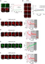
          Phospholipase C β (PLCβ), which is activated by the Gq family of heterotrimeric G proteins, hydrolyzes the inner membrane lipid phosphatidylinositol 4,5-bisphosphate (PIP2), generating diacylglycerol and inositol 1,4,5-triphosphate (IP3). Because Gq and PLCβ regulate many crucial cellular processes and have been identified as major disease drivers, activation and termination of PLCβ signaling by the Gαq subunit have been extensively studied. Gq-coupled receptor activation induces intense and transient PIP2 hydrolysis, which subsequently recovers to a low-intensity steady-state equilibrium. However, the molecular underpinnings of this equilibrium remain unclear. Here, we explored the influence of signaling crosstalk between Gq and Gi/o pathways on PIP2 metabolism in living cells using single-cell and optogenetic approaches to spatially and temporally constrain signaling. Our data suggest that the Gβγ complex is a component of the highly efficient lipase Gαq GTP–PLCβ–Gβγ. We found that over time, Gβγ dissociates from this lipase complex, leaving the less-efficient Gαq GTP–PLCβ lipase complex and allowing the significant partial recovery of PIP2 levels. Our findings also indicate that the subtype of the Gγ subunit in Gβγ fine-tunes the lipase activity of Gq–PLCβ, in which cells expressing Gγ with higher plasma membrane interaction show lower PIP2 recovery. Given that Gγ shows cell- and tissue-specific subtype expression, our findings suggest the existence of tissue-specific distinct Gq–PLCβ signaling paradigms. Furthermore, these results also outline a molecular process that likely safeguards cells from excessive Gq signaling.

          Related collections

          Most cited references79

          • Record: found
          • Abstract: found
          • Article: not found

          GTPase-activating proteins for heterotrimeric G proteins: regulators of G protein signaling (RGS) and RGS-like proteins.

          GTPase-activating proteins (GAPs) regulate heterotrimeric G proteins by increasing the rates at which their subunits hydrolyze bound GTP and thus return to the inactive state. G protein GAPs act allosterically on G subunits, in contrast to GAPs for the Ras-like monomeric GTP-binding proteins. Although they do not contribute directly to the chemistry of GTP hydrolysis, G protein GAPs can accelerate hydrolysis >2000-fold. G protein GAPs include both effector proteins (phospholipase C-¿, p115RhoGEF) and a growing family of regulators of G protein signaling (RGS proteins) that are found throughout the animal and fungal kingdoms. GAP activity can sharpen the termination of a signal upon removal of stimulus, attenuate a signal either as a feedback inhibitor or in response to a second input, promote regulatory association of other proteins, or redirect signaling within a G protein signaling network. GAPs are regulated by various controls of their cellular concentrations, by complex interactions with G¿ or with G¿5 through an endogenous G-like domain, and by interaction with multiple other proteins.
            Bookmark
            • Record: found
            • Abstract: not found
            • Article: not found

            Mean phase coherence as a measure for phase synchronization and its application to the EEG of epilepsy patients

              Bookmark
              • Record: found
              • Abstract: found
              • Article: not found

              Receptor-induced transient reduction in plasma membrane PtdIns(4,5)P2 concentration monitored in living cells.

              Although phosphatidylinositol 4,5-bisphosphate (PtdIns(4,5)P2) is a well-characterized precursor for the second messengers inositol 1,4,5-trisphosphate, diacylglycerol [1] and phosphatidylinositol 3,4,5-trisphosphate [2], it also interacts with the actin-binding proteins profilin and gelsolin [3], as well as with many signaling molecules that contain pleckstrin homology (PH) domains [4]. It is conceivable that stimuli received by receptors in the plasma membrane could be sufficiently strong to decrease the PtdIns(4,5)P2 concentration; this decrease could alter the structure of the cortical cytoskeleton and modulate the activity of signaling molecules that have PH domains. Here, we tested this hypothesis by using an in vivo fluorescent indicator for PtdIns(4,5)P2, by tagging the PH domain of phospholipase C delta 1 (PLC-delta 1) with the green fluorescent protein (GFP-PH). When expressed in cells, GFP-PH was found to be enriched at the plasma membrane. Binding studies in vitro and mutant analysis suggested that GFP-PH bound PtdIns(4,5)P2 selectively over other phosphatidylinositol lipids. Strikingly, receptor stimulation induced a transient dissociation of GFP-PH from the plasma membrane, suggesting that the concentration of PtdIns(4,5)P2 in the plasma membrane was effectively lowered. This transient dissociation was blocked by the PLC inhibitor U73122 but was not affected by the phosphoinositide (PI) 3-kinase inhibitor wortmannin, suggesting that it is mostly mediated by PLC and not by PI 3-kinase activation. Overall, our studies show that PtdIns(4,5)P2 can have second messenger functions of its own, by mediating a transient dissociation of proteins anchored in the plasma membrane.
                Bookmark

                Author and article information

                Contributors
                Journal
                J Biol Chem
                J Biol Chem
                The Journal of Biological Chemistry
                American Society for Biochemistry and Molecular Biology
                0021-9258
                1083-351X
                24 April 2021
                2021
                24 April 2021
                : 296
                : 100702
                Affiliations
                [1 ]Department of Chemistry and Biochemistry, The University of Toledo, Toledo, Ohio, USA
                [2 ]Department of Chemical Engineering, Indian Institute of Technology, Hyderabad, Sangareddy, Telangana, India
                Author notes
                []For correspondence: Ajith Karunarathne Ajith.karunarathne@ 123456utoledo.edu
                Article
                S0021-9258(21)00491-9 100702
                10.1016/j.jbc.2021.100702
                8138763
                33901492
                87f8d92d-1de0-4442-8da3-8382deebb55d
                © 2021 The Authors

                This is an open access article under the CC BY license (http://creativecommons.org/licenses/by/4.0/).

                History
                : 25 September 2020
                : 9 April 2021
                Categories
                Research Article

                Biochemistry
                plcβ,gαqgtp,pip2 recovery,gpcr,signal transduction,opsins,adaptation,optogenetics,steady-state,α2ar, α2-adrenergic receptor,β1ar, beta-1 adrenergic receptor,bopsin, blue opsin,cfp, cyan fluorescent protein,dag, diacylglycerol,dbd, dag binding domain,gap, gtpase-accelerating protein,gpcrs, g protein coupled receptors,grk3ct, g protein coupled receptor kinase 3 ct,grk-g, protein coupled receptor kinase,grprs, gastrin-releasing peptide receptors,hth, helix-turn-helix,ims, internal membranes,ip3, inositol 1,4,5-triphosphate,kor, κ-opioid receptor,m1r, m1-muscarinic receptor,m3r, m3-muscarinic receptor,mch–ph, mcherry–ph,mgq, mini gq protein,ne, norepinephrine,pd, phase difference,phlp, phosducin-like protein,ph, pleckstrin homology,pi4, phosphatidylinositol 4,pip2, phosphatidylinositol 4,5-bisphosphate,pip3, phosphatidylinositol (3,4,5)-trisphosphate,plc, phospholipase c,plcβ, phospholipase c β,pm, plasma membrane,ptx, pertussis toxin,rgs, regulators of g protein signaling,t1/2, halftime,yfp, yellow fluorescent protein

                Comments

                Comment on this article