30
views
0
recommends
+1 Recommend
0 collections
    0
    shares
      • Record: found
      • Abstract: found
      • Article: found
      Is Open Access

      Variability of Foveal Avascular Zone Metrics Derived From Optical Coherence Tomography Angiography Images

      research-article

      Read this article at

      Bookmark
          There is no author summary for this article yet. Authors can add summaries to their articles on ScienceOpen to make them more accessible to a non-specialist audience.

          Abstract

          Purpose

          To characterize sources of inter- and intrasubject variability in quantitative foveal avascular zone (FAZ) metrics.

          Methods

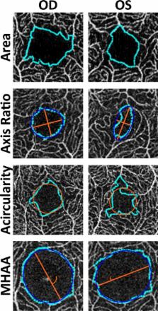
          Two 3×3-mm optical coherence tomography angiography scans (centered on the fovea) were acquired in both eyes of 175 subjects. An image of the superficial plexus was extracted from each scan and segmented twice by a single observer. Four quantitative FAZ morphology metrics (area, axis ratio, acircularity, major horizontal axis angle) were calculated, and a variance components analysis was performed.

          Results

          Mean (±SD) age was 27.9 ± 11.9 years, and 55% were female. Area had the largest amount of variance resulting from intersubject differences (93.1%). In contrast, there was large interocular variance for axis ratio, acircularity, and major horizontal axis angle (55.0%, 53.7%, 70.7%, respectively), though only axis ratio showed significant asymmetry between fellow eyes ( P < 0.05). Neither repeated images from the same eye nor repeated segmentation on the same image were significant sources of variance.

          Conclusions

          Metrics of FAZ morphology show excellent repeatability and reliability. Excluding FAZ area, there was a high amount of variance attributed to interocular differences for the other FAZ metrics; therefore, the fellow eye should not be considered a control for FAZ studies when using these metrics.

          Translational Relevance

          Vision scientists must be prudent when choosing FAZ metrics, as they display varying degrees of within-subject differences relative to between-subject differences. It seems likely that different metrics will be best suited for different tasks, such as monitoring small changes over time within a single subject or assessing whether a given FAZ is abnormal.

          Related collections

          Most cited references51

          • Record: found
          • Abstract: found
          • Article: not found

          NIH Image to ImageJ: 25 years of image analysis.

          For the past 25 years NIH Image and ImageJ software have been pioneers as open tools for the analysis of scientific images. We discuss the origins, challenges and solutions of these two programs, and how their history can serve to advise and inform other software projects.
            Bookmark
            • Record: found
            • Abstract: found
            • Article: not found

            ENLARGEMENT OF FOVEAL AVASCULAR ZONE IN DIABETIC EYES EVALUATED BY EN FACE OPTICAL COHERENCE TOMOGRAPHY ANGIOGRAPHY.

            To evaluate the area of the foveal avascular zone (FAZ) detected by en face OCTA (AngioVue, Avanti OCT; Optovue) in healthy and diabetic eyes.
              Bookmark
              • Record: found
              • Abstract: found
              • Article: not found

              CORRELATION OF FOVEAL AVASCULAR ZONE SIZE WITH FOVEAL MORPHOLOGY IN NORMAL EYES USING OPTICAL COHERENCE TOMOGRAPHY ANGIOGRAPHY.

              To analyze the foveal avascular zone (FAZ) in normal eyes using optical coherence tomography angiography.
                Bookmark

                Author and article information

                Journal
                Transl Vis Sci Technol
                Transl Vis Sci Technol
                tvst
                Transl Vis Sci Technol
                TVST
                Translational Vision Science & Technology
                The Association for Research in Vision and Ophthalmology
                2164-2591
                September 2018
                1 October 2018
                : 7
                : 5
                : 20
                Affiliations
                [1 ]Cell Biology, Neurobiology, & Anatomy, Medical College of Wisconsin, Milwaukee, WI, USA
                [2 ]National Institute for Health Research Biomedical Research Centre, Moorfields Eye Hospital and UCL Institute of Ophthalmology, London, UK
                [3 ]Ophthalmology & Visual Sciences, Medical College of Wisconsin, Milwaukee, WI, USA
                [4 ]School of Medicine, Medical College of Wisconsin, Milwaukee, WI, USA
                [5 ]Division of Biostatistics, Institute of Health and Equity, Medical College of Wisconsin, Milwaukee, WI, USA
                Author notes
                Correspondence: Joseph Carroll, Department of Ophthalmology & Visual Sciences, Medical College of Wisconsin, 925 N. 87th Street, Milwaukee, WI 53226-0509, USA; email: jcarroll@ 123456mcw.edu
                Article
                tvst-07-05-07 TVST-18-0907
                10.1167/tvst.7.5.20
                6166903
                30280005
                8a23e27b-b4e1-4dc9-a7f9-f781c3492cb7
                Copyright 2018 The Authors

                This work is licensed under a Creative Commons Attribution-NonCommercial-NoDerivatives 4.0 International License.

                History
                : 30 May 2018
                : 10 August 2018
                Categories
                Articles

                foveal avascular zone,imaging,optical coherence tomography angiography,foveal morphology,variability,acircularity,axis ratio

                Comments

                Comment on this article