29
views
0
recommends
+1 Recommend
0 collections
    0
    shares
      • Record: found
      • Abstract: found
      • Article: found
      Is Open Access

      Revisiting in vivo staining with alizarin red S - a valuable approach to analyse zebrafish skeletal mineralization during development and regeneration

      research-article

      Read this article at

      Bookmark
          There is no author summary for this article yet. Authors can add summaries to their articles on ScienceOpen to make them more accessible to a non-specialist audience.

          Abstract

          Background

          The correct evaluation of mineralization is fundamental for the study of skeletal development, maintenance, and regeneration. Current methods to visualize mineralized tissue in zebrafish rely on: 1) fixed specimens; 2) radiographic and μCT techniques, that are ultimately limited in resolution; or 3) vital stains with fluorochromes that are indistinguishable from the signal of green fluorescent protein (GFP)-labelled cells. Alizarin compounds, either in the form of alizarin red S (ARS) or alizarin complexone (ALC), have long been used to stain the mineralized skeleton in fixed specimens from all vertebrate groups. Recent works have used ARS vital staining in zebrafish and medaka, yet not based on consistent protocols. There is a fundamental concern on whether ARS vital staining, achieved by adding ARS to the water, can affect bone formation in juvenile and adult zebrafish, as ARS has been shown to inhibit skeletal growth and mineralization in mammals.

          Results

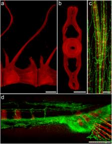
          Here we present a protocol for vital staining of mineralized structures in zebrafish with a low ARS concentration that does not affect bone mineralization, even after repetitive ARS staining events, as confirmed by careful imaging under fluorescent light. Early and late stages of bone development are equally unaffected by this vital staining protocol. From all tested concentrations, 0.01 % ARS yielded correct detection of bone calcium deposits without inducing additional stress to fish.

          Conclusions

          The proposed ARS vital staining protocol can be combined with GFP fluorescence associated with skeletal tissues and thus represents a powerful tool for in vivo monitoring of mineralized structures. We provide examples from wild type and transgenic GFP-expressing zebrafish, for endoskeletal development and dermal fin ray regeneration.

          Related collections

          Most cited references69

          • Record: found
          • Abstract: not found
          • Article: not found

          Bone histomorphometry: standardization of nomenclature, symbols, and units. Report of the ASBMR Histomorphometry Nomenclature Committee.

            Bookmark
            • Record: found
            • Abstract: found
            • Article: not found

            The identification of genes with unique and essential functions in the development of the zebrafish, Danio rerio.

            In a large-scale screen, we isolated mutants displaying a specific visible phenotype in embryos or early larvae of the zebrafish, Danio rerio. Males were mutagenized with ethylnitrosourea (ENU) and F2 families of single pair matings between sibling F1 fish, heterozygous for a mutagenized genome, were raised. Egg lays were obtained from several crosses between F2 siblings, resulting in scoring of 3857 mutagenized genomes. F3 progeny were scored at the second, third and sixth day of development, using a stereomicroscope. In a subsequent screen, fixed embryos were analyzed for correct retinotectal projection. A total of 4264 mutants were identified. Two thirds of the mutants displaying rather general abnormalities were eventually discarded. We kept and characterized 1163 mutants. In complementation crosses performed between mutants with similar phenotypes, 894 mutants have been assigned to 372 genes. The average allele frequency is 2.4. We identified genes involved in early development, notochord, brain, spinal cord, somites, muscles, heart, circulation, blood, skin, fin, eye, otic vesicle, jaw and branchial arches, pigment pattern, pigment formation, gut, liver, motility and touch response. Our collection contains alleles of almost all previously described zebrafish mutants. From the allele frequencies and other considerations we estimate that the 372 genes defined by the mutants probably represent more than half of all genes that could have been discovered using the criteria of our screen. Here we give an overview of the spectrum of mutant phenotypes obtained, and discuss the limits and the potentials of a genetic saturation screen in the zebrafish.
              Bookmark
              • Record: found
              • Abstract: found
              • Article: not found

              A two-color acid-free cartilage and bone stain for zebrafish larvae.

              Traditionally, cartilage is stained by alcian blue using acidic conditions to differentiate tissue staining. The acidic conditions are problematic when one wishes to stain the same specimen for mineralized bone with alizarin red, because acid demineralizes bone, which negatively affects bone staining. We have developed an acid-free method to stain cartilage and bone simultaneously in zebrafish larvae. This method has the additional advantage that PCR genotyping of stained specimens is possible.
                Bookmark

                Author and article information

                Contributors
                bensimonbrito@gmail.com
                jpgsilva@ualg.pt
                gisela.dionisio@ua.pt
                ann.huysseune@ugent.be
                lcancela@ualg.pt
                PEckhardWitten@aol.com
                Journal
                BMC Dev Biol
                BMC Dev. Biol
                BMC Developmental Biology
                BioMed Central (London )
                1471-213X
                19 January 2016
                19 January 2016
                2016
                : 16
                : 2
                Affiliations
                [ ]Centre of Marine Sciences – CCMar, University of Algarve, Campus de Gambelas, Faro, Portugal
                [ ]Evolutionary Developmental Biology, Biology Department, Ghent University, Ghent, Belgium
                [ ]ProRegeM PhD Programme, Department of Biomedical Sciences and Medicine, University of Algarve, Campus de Gambelas, Faro, Portugal
                [ ]Guia Marine Laboratory, Oceanography Centre, Faculty of Sciences of University of Lisbon, Cascais, Portugal
                [ ]Department of Biomedical Sciences and Medicine, University of Algarve, Campus de Gambelas, Faro, Portugal
                [ ]Current address: CEDOC - Faculdade de Ciências Médicas, Universidade Nova de Lisboa, Lisbon, Portugal
                Author information
                http://orcid.org/0000-0003-1663-2232
                Article
                102
                10.1186/s12861-016-0102-4
                4719692
                26787303
                8a4ff3db-1c6a-4278-bd4b-c827eb1a256f
                © Bensimon-Brito et al. 2016

                Open AccessThis article is distributed under the terms of the Creative Commons Attribution 4.0 International License ( http://creativecommons.org/licenses/by/4.0/), which permits unrestricted use, distribution, and reproduction in any medium, provided you give appropriate credit to the original author(s) and the source, provide a link to the Creative Commons license, and indicate if changes were made. The Creative Commons Public Domain Dedication waiver ( http://creativecommons.org/publicdomain/zero/1.0/) applies to the data made available in this article, unless otherwise stated.

                History
                : 24 August 2015
                : 8 January 2016
                Funding
                Funded by: FundRef http://dx.doi.org/10.13039/501100001871, Fundação para a Ciência e a Tecnologia (PT);
                Award ID: PEst-C/MAR/LA0015/2011
                Award Recipient :
                Funded by: Fonds Voor Wetenschappelijk Onderzoek
                Award ID: FWO 3G.0040.08
                Award Recipient :
                Categories
                Methodology Article
                Custom metadata
                © The Author(s) 2016

                Developmental biology
                vertebral column,caudal fin,mineral apposition,bone,fluorescence imaging,calcium,hydroxyapatite,alizarin red s

                Comments

                Comment on this article