7
views
0
recommends
+1 Recommend
0 collections
    0
    shares
      • Record: found
      • Abstract: found
      • Article: found
      Is Open Access

      Differential functional connectivity underlying asymmetric reward-related activity in human and nonhuman primates

      research-article

      Read this article at

      Bookmark
          There is no author summary for this article yet. Authors can add summaries to their articles on ScienceOpen to make them more accessible to a non-specialist audience.

          Significance

          Lateralization of functions in the brain has been demonstrated in many different cognitive processes. It is supposed to increase processing abilities by reducing bilateral redundancy. Yet lateralization of reward processing, despite extremely common asymmetrical findings, has received little attention. Our neuroimaging study shows a functional lateralization of the response to reward in the lateral part of the orbitofrontal cortex (OFC), together with an asymmetric functional connectivity pattern. This particular feature was identified not only in humans but also in nonhuman primates. Our findings challenge the classical view of the OFC as a symmetrical brain region. They are urging the need for considering the specific contribution of the left and right OFC when investigating reward-related signals.

          Abstract

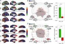
          The orbitofrontal cortex (OFC) is a key brain region involved in complex cognitive functions such as reward processing and decision making. Neuroimaging studies have reported unilateral OFC response to reward-related variables; however, those studies rarely discussed this observation. Nevertheless, some lesion studies suggest that the left and right OFC contribute differently to cognitive processes. We hypothesized that the OFC asymmetrical response to reward could reflect underlying hemispherical difference in OFC functional connectivity. Using resting-state and reward-related functional MRI data from humans and from rhesus macaques, we first identified an asymmetrical response of the lateral OFC to reward in both species. Crucially, the subregion showing the highest reward-related asymmetry (RRA) overlapped with the region showing the highest functional connectivity asymmetry (FCA). Furthermore, the two types of asymmetries were found to be significantly correlated across individuals. In both species, the right lateral OFC was more connected to the default mode network compared to the left lateral OFC. Altogether, our results suggest a functional specialization of the left and right lateral OFC in primates.

          Related collections

          Most cited references85

          • Record: found
          • Abstract: found
          • Article: not found

          FSL.

          FSL (the FMRIB Software Library) is a comprehensive library of analysis tools for functional, structural and diffusion MRI brain imaging data, written mainly by members of the Analysis Group, FMRIB, Oxford. For this NeuroImage special issue on "20 years of fMRI" we have been asked to write about the history, developments and current status of FSL. We also include some descriptions of parts of FSL that are not well covered in the existing literature. We hope that some of this content might be of interest to users of FSL, and also maybe to new research groups considering creating, releasing and supporting new software packages for brain image analysis. Copyright © 2011 Elsevier Inc. All rights reserved.
            Bookmark
            • Record: found
            • Abstract: found
            • Article: not found

            FreeSurfer.

            FreeSurfer is a suite of tools for the analysis of neuroimaging data that provides an array of algorithms to quantify the functional, connectional and structural properties of the human brain. It has evolved from a package primarily aimed at generating surface representations of the cerebral cortex into one that automatically creates models of most macroscopically visible structures in the human brain given any reasonable T1-weighted input image. It is freely available, runs on a wide variety of hardware and software platforms, and is open source. Copyright © 2012 Elsevier Inc. All rights reserved.
              Bookmark
              • Record: found
              • Abstract: found
              • Article: not found

              Advances in functional and structural MR image analysis and implementation as FSL.

              The techniques available for the interrogation and analysis of neuroimaging data have a large influence in determining the flexibility, sensitivity, and scope of neuroimaging experiments. The development of such methodologies has allowed investigators to address scientific questions that could not previously be answered and, as such, has become an important research area in its own right. In this paper, we present a review of the research carried out by the Analysis Group at the Oxford Centre for Functional MRI of the Brain (FMRIB). This research has focussed on the development of new methodologies for the analysis of both structural and functional magnetic resonance imaging data. The majority of the research laid out in this paper has been implemented as freely available software tools within FMRIB's Software Library (FSL).
                Bookmark

                Author and article information

                Journal
                Proc Natl Acad Sci U S A
                Proc Natl Acad Sci U S A
                pnas
                pnas
                PNAS
                Proceedings of the National Academy of Sciences of the United States of America
                National Academy of Sciences
                0027-8424
                1091-6490
                10 November 2020
                29 October 2020
                29 October 2020
                : 117
                : 45
                : 28452-28462
                Affiliations
                [1] aWellcome Integrative Neuroimaging Centre, Department of Experimental Psychology, University of Oxford , Oxford OX2 6GG, United Kingdom;
                [2] bFrontal Function and Pathology team, Institut du Cerveau, Sorbonne Université, INSERM U 1127, CNRS UMR 7225, 75013 Paris, France;
                [3] cSchool of Psychology, University of Plymouth , Plymouth PL4 8AA, United Kingdom;
                [4] dStem Cell and Brain Research Institute, INSERM U 1208, Université Lyon 1 , 69500 Bron, France
                Author notes
                1To whom correspondence may be addressed. Email: lopez.alizee@ 123456gmail.com or jerome.sallet@ 123456psy.ox.ac.uk .

                Edited by Peter H. Rudebeck, Icahn School of Medicine at Mount Sinai, New York, NY, and accepted by Editorial Board Member Leslie G. Ungerleider September 16, 2020 (received for review January 17, 2020)

                Author contributions: A.L.-P. and J.S. designed research; A.L.-P., L.R., D.F., K.M., E.F.F., N.K., M.F.S.R., and J.S. performed research; A.L.-P. analyzed data; and A.L.-P., M.F.S.R., and J.S. wrote the paper.

                Author information
                https://orcid.org/0000-0002-7566-5715
                https://orcid.org/0000-0003-2195-7621
                https://orcid.org/0000-0003-1653-5969
                https://orcid.org/0000-0003-0663-5253
                https://orcid.org/0000-0003-1485-0332
                https://orcid.org/0000-0002-7878-0209
                Article
                202000759
                10.1073/pnas.2000759117
                7668182
                33122437
                8c12d99b-7fe0-4577-80bf-c5d157d7469f
                Copyright © 2020 the Author(s). Published by PNAS.

                This open access article is distributed under Creative Commons Attribution License 4.0 (CC BY).

                History
                Page count
                Pages: 11
                Funding
                Funded by: Wellcome 100010269
                Award ID: 203139/Z/16/Z
                Award Recipient : Matthew F.S. Rushworth Award Recipient : Jerome Sallet
                Funded by: Wellcome 100010269
                Award ID: WT100973AIA
                Award Recipient : Matthew F.S. Rushworth Award Recipient : Jerome Sallet
                Funded by: Wellcome 100010269
                Award ID: 103184/Z/13/Z
                Award Recipient : Matthew F.S. Rushworth Award Recipient : Jerome Sallet
                Funded by: Wellcome 100010269
                Award ID: 202831/Z/16/Z
                Award Recipient : Matthew F.S. Rushworth Award Recipient : Jerome Sallet
                Funded by: Wellcome 100010269
                Award ID: 105238/Z/14/Z
                Award Recipient : Matthew F.S. Rushworth Award Recipient : Jerome Sallet
                Funded by: RCUK | Medical Research Council (MRC) 501100000265
                Award ID: MR/P024955/1
                Award Recipient : Matthew F.S. Rushworth Award Recipient : Jerome Sallet
                Funded by: RCUK | Medical Research Council (MRC) 501100000265
                Award ID: G0902373
                Award Recipient : Matthew F.S. Rushworth Award Recipient : Jerome Sallet
                Funded by: Fondation Bettencourt Schueller (Bettencourt Schueller Foundation) 501100007492
                Award ID: NA
                Award Recipient : Matthew F.S. Rushworth Award Recipient : Jerome Sallet
                Categories
                Biological Sciences
                Neuroscience

                orbitofrontal cortex,reward,functional connectivity,lateralization

                Comments

                Comment on this article