1
views
0
recommends
+1 Recommend
0 collections
    0
    shares
      • Record: found
      • Abstract: found
      • Article: found
      Is Open Access

      Neoplastic lesions in domestic pigs detected at slaughter: literature review and a 20-year review (1998–2018) of carcass inspection in Catalonia

      research-article

      Read this article at

      Bookmark
          There is no author summary for this article yet. Authors can add summaries to their articles on ScienceOpen to make them more accessible to a non-specialist audience.

          Abstract

          Background

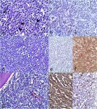
          The present paper reviews the occurrence of neoplasms in swine and presents a case series of 56 tumors submitted to the Slaughterhouse Support Network ( Servei de Suport a Escorxadors [SESC] IRTA-CReSA]) from slaughtered pigs from 1998 to 2018 (April) in Catalonia (Spain). The aim of the study was to describe the spectrum of spontaneous neoplastic lesions found in slaughtered pigs and to compare the reported tumor cases with previous published data. Lymphoid neoplasms were characterized and classified using the WHO classification adapted for animals.

          Results

          The most reported neoplasm during this period was lymphoma (28). Within lymphomas, the B-cell type was the most common, being the diffuse large B-cell lymphoma (15/28) the most represented subtype. Other submitted non-lymphoid neoplasms included melanoma (7), nephroblastoma (3), mast cell tumor (2), liposarcoma (2), osteochondromatosis (2), papillary cystadenocarcinoma (1), peripheral nerve sheath tumor (1), lymphoid leukemia (1), fibropapilloma (1), hemangiosarcoma (1), hepatoma (1), histiocytic sarcoma (1), pheochromocytoma (1) and osteosarcoma (1).

          Conclusions

          The existence of a well-established Slaughterhouse Support Network allowed the compilation of comprehensive data for further epidemiological and pathological studies, particularly about less commonly reported lesions in livestock such as neoplasms in pigs.

          Supplementary Information

          The online version contains supplementary material available at 10.1186/s40813-021-00207-0.

          Related collections

          Most cited references84

          • Record: found
          • Abstract: found
          • Article: not found

          Classification of canine malignant lymphomas according to the World Health Organization criteria.

          A study was carried out to test the accuracy and consistency of veterinary pathologists, not specialists in hematopathology, in applying the World Health Organization (WHO) system of classification of canine lymphomas. This study represents an initiative of the ACVP Oncology Committee, and the classification has been endorsed by the World Small Animal Veterinary Association (WASVA). Tissue biopsies from cases of canine lymphoma were received from veterinary oncologists, and a study by pathologists given only signalment was carried out on 300 cases. Twenty pathologists reviewed these 300 cases with each required to choose a diagnosis from a list of 43 B and T cell lymphomas. Three of the 20 were hematopathologists who determined the consensus diagnosis for each case. The 17 who formed the test group were experienced but not specialists in hematopathology, and most were diplomates of the American or European Colleges of Veterinary Pathology. The overall accuracy of the 17 pathologists on the 300 cases was 83%. When the analysis was limited to the 6 most common diagnoses, containing 80% of all cases, accuracy rose to 87%. In a test of reproducibility enabled by reintroducing 5% of cases entered under a different identity, the overall agreement between the first and second diagnosis ranged from 40 to 87%. The statistical review included 43,000 data points for each of the 20 pathologists.
            Bookmark
            • Record: found
            • Abstract: found
            • Article: not found

            The pig as an animal model for human pathologies: A proteomics perspective.

            Traditional biomedical models are easy to manage in experimental facilities and allow fast and affordable basic genetic studies related to human disorders, but in some cases they do not always represent the complexity of their physiology. Translational medicine demands selected models depending on the particularities of the human disease to be investigated, reproducing as closely as possible the evolution, clinical symptoms and molecular pathways, cells or tissues involved in the dysfunction. Thus, pig models offer an alternative because of their anatomical and physiological similarities to humans and the availability of genomic, transcriptomic and, progressively more, proteomic tools for analysis of this species. Furthermore, there is a wide range of natural, selected and transgenic porcine breeds. The present review provides a summary of the applications of the pig as a model for metabolic, cardiovascular, infectious diseases, xenotransplantation and neurological disorders and an overview of the possibilities that the diverse proteomic techniques offer to study these pathologies in depth.
              Bookmark
              • Record: found
              • Abstract: found
              • Article: found
              Is Open Access

              The pig as a model for translational research: overview of porcine animal models at Jichi Medical University

              To improve the welfare of experimental animals, investigators seek to respect the 3R principle (Replacement, Reduction, and Refinement). Even when large animal studies are essential before moving to clinical trials, it is important to look for ways to reduce the number of experimental animals used. At the Center for the Development of Advanced Medical Technology, we consider ‘medical’ pigs to be ideal preclinical model systems. We have been using both wild-type and genetically modified pigs. We began using this approach about 10 years ago with a ‘total pig system’ to model human health and disease for the purposes of both medical skill education and the development of new devices and therapeutic strategies. At our Center, medical students and residents use pigs to gain experience with surgical skills and train for emergency procedures after appropriate simulation training. Senior clinicians have also used these models to advance the development of innovative tools for endo- and laparoscopic procedures. The Center focuses on translational research for organ transplantation and stem cell therapy. Several pig models have been established for liver, intestine, kidney, pancreas, and lung transplantation. Mesenchymal stromal cells have been established in green fluorescent protein- and red fluorescent protein-transgenic pigs and tested to trans-differentiate organogenesis. A program to establish induced pluripotent stem cells in the pig is ongoing at our Center. Here, we review our 10 years of activity in this field. Based on our experience in surgical education and research, experimental pigs are valuable models in translational research.
                Bookmark

                Author and article information

                Contributors
                enric.vidal@irta.cat
                Journal
                Porcine Health Manag
                Porcine Health Manag
                Porcine Health Management
                BioMed Central (London )
                2055-5660
                7 April 2021
                7 April 2021
                2021
                : 7
                : 30
                Affiliations
                [1 ]GRID grid.7080.f, Servei de Diagnòstic de Patologia Veterinària (SDPV), Departament de Sanitat i d’Anatomia Animals, , Universitat Autonòma de Barcelona, ; 08193 Bellaterra (Cerdanyola del Vallès), Barcelona, Spain
                [2 ]GRID grid.7080.f, IRTA, Centre de Recerca en Sanitat Animal (CReSA, IRTA-UAB), , Campus de la Universitat Autònoma de Barcelona, ; 08193 Bellaterra (Cerdanyola del Vallès), Barcelona Spain
                Author information
                http://orcid.org/0000-0002-4965-3286
                Article
                207
                10.1186/s40813-021-00207-0
                8025367
                8d42ec8c-396c-4d67-ad04-aa4f1e286a5e
                © The Author(s) 2021

                Open AccessThis article is licensed under a Creative Commons Attribution 4.0 International License, which permits use, sharing, adaptation, distribution and reproduction in any medium or format, as long as you give appropriate credit to the original author(s) and the source, provide a link to the Creative Commons licence, and indicate if changes were made. The images or other third party material in this article are included in the article's Creative Commons licence, unless indicated otherwise in a credit line to the material. If material is not included in the article's Creative Commons licence and your intended use is not permitted by statutory regulation or exceeds the permitted use, you will need to obtain permission directly from the copyright holder. To view a copy of this licence, visit http://creativecommons.org/licenses/by/4.0/. The Creative Commons Public Domain Dedication waiver ( http://creativecommons.org/publicdomain/zero/1.0/) applies to the data made available in this article, unless otherwise stated in a credit line to the data.

                History
                : 15 February 2021
                : 22 March 2021
                Categories
                Research
                Custom metadata
                © The Author(s) 2021

                tumor,neoplasm,slaughterhouse,sus scrofa,swine,food inspection,surveillance,lymphoma,retrospective study

                Comments

                Comment on this article