36
views
0
recommends
+1 Recommend
0 collections
    0
    shares
      • Record: found
      • Abstract: found
      • Article: found
      Is Open Access

      chaoptin, prominin, eyes shut and crumbs form a genetic network controlling the apical compartment of Drosophila photoreceptor cells

      research-article

      Read this article at

      Bookmark
          There is no author summary for this article yet. Authors can add summaries to their articles on ScienceOpen to make them more accessible to a non-specialist audience.

          ABSTRACT

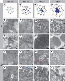
          The apical surface of epithelial cells is often highly specialised to fulfil cell type-specific functions. Many epithelial cells expand their apical surface by forming microvilli, actin-based, finger-like membrane protrusions. The apical surface of Drosophila photoreceptor cells (PRCs) forms tightly packed microvilli, which are organised into the photosensitive rhabdomeres. As previously shown, the GPI-anchored adhesion protein Chaoptin is required for the stability of the microvilli, whereas the transmembrane protein Crumbs is essential for proper rhabdomere morphogenesis. Here we show that chaoptin synergises with crumbs to ensure optimal rhabdomere width. In addition, reduction of crumbs ameliorates morphogenetic defects observed in PRCs mutant for prominin and eyes shut, known antagonists of chaoptin. These results suggest that these four genes provide a balance of adhesion and anti-adhesion to maintain microvilli development and maintenance. Similar to crumbs mutant PRCs, PRCs devoid of prominin or eyes shut undergo light-dependent retinal degeneration. Given the observation that human orthologues of crumbs, prominin and eyes shut result in progressive retinal degeneration and blindness, the Drosophila eye is ideally suited to unravel the genetic and cellular mechanisms that ensure morphogenesis of PRCs and their maintenance under light-mediated stress.

          Related collections

          Most cited references70

          • Record: found
          • Abstract: found
          • Article: not found

          From the Cover: Directed, efficient, and versatile modifications of the Drosophila genome by genomic engineering.

          With the completion of genome sequences of major model organisms, increasingly sophisticated genetic tools are necessary for investigating the complex and coordinated functions of genes. Here we describe a genetic manipulation system termed "genomic engineering" in Drosophila. Genomic engineering is a 2-step process that combines the ends-out (replacement) gene targeting with phage integrase phiC31-mediated DNA integration. First, through an improved and modified gene targeting method, a founder knock-out line is generated by deleting the target gene and replacing it with an integration site of phiC31. Second, DNA integration by phiC31 is used to reintroduce modified target-gene DNA into the native locus in the founder knock-out line. Genomic engineering permits directed and highly efficient modifications of a chosen genomic locus into virtually any desired mutant allele. We have successfully applied the genomic engineering scheme on 6 different genes and have generated at their loci more than 70 unique alleles.
            Bookmark
            • Record: found
            • Abstract: found
            • Article: not found

            Mutations in a human homologue of Drosophila crumbs cause retinitis pigmentosa (RP12).

            Retinitis pigmentosa (RP) comprises a clinically and genetically heterogeneous group of diseases that afflicts approximately 1.5 million people worldwide. Affected individuals suffer from a progressive degeneration of the photoreceptors, eventually resulting in severe visual impairment. To isolate candidate genes for chorioretinal diseases, we cloned cDNAs specifically or preferentially expressed in the human retina and the retinal pigment epithelium (RPE) through a novel suppression subtractive hybridization (SSH) method. One of these cDNAs (RET3C11) mapped to chromosome 1q31-q32.1, a region harbouring a gene involved in a severe form of autosomal recessive RP characterized by a typical preservation of the para-arteriolar RPE (RP12; ref. 3). The full-length cDNA encodes an extracellular protein with 19 EGF-like domains, 3 laminin A G-like domains and a C-type lectin domain. This protein is homologous to the Drosophila melanogaster protein crumbs (CRB), and denoted CRB1 (crumbs homologue 1). In ten unrelated RP patients with preserved para-arteriolar RPE, we identified a homozygous AluY insertion disrupting the ORF, five homozygous missense mutations and four compound heterozygous mutations in CRB1. The similarity to CRB suggests a role for CRB1 in cell-cell interaction and possibly in the maintenance of cell polarity in the retina. The distinct RPE abnormalities observed in RP12 patients suggest that CRB1 mutations trigger a novel mechanism of photoreceptor degeneration.
              Bookmark
              • Record: found
              • Abstract: found
              • Article: not found

              Crumbs, the Drosophila homologue of human CRB1/RP12, is essential for photoreceptor morphogenesis.

              The apical transmembrane protein Crumbs is a central regulator of epithelial apical-basal polarity in Drosophila. Loss-of-function mutations in the human homologue of Crumbs, CRB1 (RP12), cause recessive retinal dystrophies, including retinitis pigmentosa. Here we show that Crumbs and CRB1 localize to corresponding subdomains of the photoreceptor apical plasma membrane: the stalk of the Drosophila photoreceptor and the inner segment of mammalian photoreceptors. These subdomains support the morphogenesis and orientation of the photosensitive membrane organelles: rhabdomeres and outer segments, respectively. Drosophila Crumbs is required to maintain zonula adherens integrity during the rapid apical membrane expansion that builds the rhabdomere. Crumbs also regulates stalk development by stabilizing the membrane-associated spectrin cytoskeleton, a function mechanistically distinct from its role in epithelial apical-basal polarity. We propose that Crumbs is a central component of a molecular scaffold that controls zonula adherens assembly and defines the stalk as an apical membrane subdomain. Defects in such scaffolds may contribute to human CRB1-related retinal dystrophies.
                Bookmark

                Author and article information

                Journal
                Biol Open
                Biol Open
                biolopen
                bio
                Biology Open
                The Company of Biologists (Bidder Building, 140 Cowley Road, Cambridge, CB4 0DL, UK )
                2046-6390
                15 May 2014
                4 April 2014
                : 3
                : 5
                : 332-341
                Affiliations
                [1]Max Planck Institute of Molecular Cell Biology and Genetics , Pfotenhauerstrasse 108, D-01307 Dresden, Germany
                Author notes
                [* ]Author for correspondence ( knust@ 123456mpi-cbg.de )
                Article
                BIO20147310
                10.1242/bio.20147310
                4021355
                24705015
                8e7a2a36-0ed6-47b6-a458-d042fb57bcc9
                © 2014. Published by The Company of Biologists Ltd

                This is an Open Access article distributed under the terms of the Creative Commons Attribution License ( http://creativecommons.org/licenses/by/3.0), which permits unrestricted use, distribution and reproduction in any medium provided that the original work is properly attributed.

                History
                : 3 December 2013
                : 27 February 2014
                Categories
                Research Article

                Life sciences
                microvilli,retinal degeneration,rhabdomere,adhesion
                Life sciences
                microvilli, retinal degeneration, rhabdomere, adhesion

                Comments

                Comment on this article