43
views
0
recommends
+1 Recommend
0 collections
    0
    shares
      • Record: found
      • Abstract: found
      • Article: not found

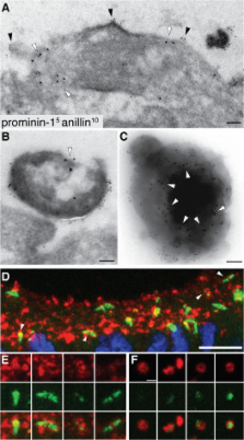
      Midbody and primary cilium of neural progenitors release extracellular membrane particles enriched in the stem cell marker prominin-1

      research-article

      Read this article at

      ScienceOpenPublisherPMC
      Bookmark
          There is no author summary for this article yet. Authors can add summaries to their articles on ScienceOpen to make them more accessible to a non-specialist audience.

          Abstract

          Expansion of the neocortex requires symmetric divisions of neuroepithelial cells, the primary progenitor cells of the developing mammalian central nervous system. Symmetrically dividing neuroepithelial cells are known to form a midbody at their apical (rather than lateral) surface. We show that apical midbodies of neuroepithelial cells concentrate prominin-1 (CD133), a somatic stem cell marker and defining constituent of a specific plasma membrane microdomain. Moreover, these apical midbodies are released, as a whole or in part, into the extracellular space, yielding the prominin-1–enriched membrane particles found in the neural tube fluid. The primary cilium of neuroepithelial cells also concentrates prominin-1 and appears to be a second source of the prominin-1–bearing extracellular membrane particles. Our data reveal novel origins of extracellular membrane traffic that enable neural stem and progenitor cells to avoid the asymmetric inheritance of the midbody observed for other cells and, by releasing a stem cell membrane microdomain, to potentially influence the balance of their proliferation versus differentiation.

          Related collections

          Most cited references53

          • Record: found
          • Abstract: found
          • Article: not found

          RADIOAUTOGRAPHIC STUDIES OF CHOLINE INCORPORATION INTO PERIPHERAL NERVE MYELIN

          This radioautographic study was designed to localize the cytological sites involved in the incorporation of a lipid precursor into the myelin and the myelin-related cell of the peripheral nervous system. Both myelinating and fully myelinated cultures of rat dorsal root ganglia were exposed to a 30-min pulse of tritiated choline and either fixed immediately or allowed 6 or 48 hr of chase incubation before fixation. After Epon embedding, light and electron microscopic radioautograms were prepared with Ilford L-4 emulsion. Analysis of the pattern of choline incorporation into myelinating cultures indicated that radioactivity appeared all along the length of the internode, without there being a preferential site of initial incorporation. Light microscopic radioautograms of cultures at varying states of maturity were compared in order to determine the relative degree of myelin labeling. This analysis indicated that the myelin-Schwann cell unit in the fully myelinated cultures incorporated choline as actively as did this unit in the myelinating cultures. Because of technical difficulties, it was not possible to determine the precise localization of the incorporated radioactivity within the compact myelin. These data are related to recent biochemical studies indicating that the mature myelin of the central nervous system does incorporate a significant amount of lipid precursor under the appropriate experimental conditions. These observations support the concept that a significant amount of myelin-related metabolic activity occurs in mature tissue; this activity is considered part of an essential and continuous process of myelin maintenance and repair.
            Bookmark
            • Record: found
            • Abstract: not found
            • Article: not found

            The cell biology of neurogenesis.

            During the development of the mammalian central nervous system, neural stem cells and their derivative progenitor cells generate neurons by asymmetric and symmetric divisions. The proliferation versus differentiation of these cells and the type of division are closely linked to their epithelial characteristics, notably, their apical-basal polarity and cell-cycle length. Here, we discuss how these features change during development from neuroepithelial to radial glial cells, and how this transition affects cell fate and neurogenesis.
              Bookmark
              • Record: found
              • Abstract: found
              • Article: not found

              Cilia and Hedgehog responsiveness in the mouse.

              The intraflagellar transport (IFT) proteins Ift172/Wimple and Polaris/Ift88 and the anterograde IFT motor kinesin-II are required for the production and maintenance of cilia. These proteins are also required for the activation of targets of the mouse Hedgehog (Hh) pathway by Gli transcription factors. The phenotypes of the IFT mutants, however, are not identical to mutants that lack Smoothened (Smo), an essential activator of the Hh pathway. We show here that mouse embryos that lack both Ift172 and Smo are identical to Ift172 single mutants, which indicates that Ift172 acts downstream of Smo. Ift172 mutants have a weaker neural patterning phenotype than Smo mutants, because Ift172, but not Smo, is required for proteolytic processing of Gli3 to its repressor form. Dnchc2 and Kif3a, essential subunits of the retrograde and anterograde IFT motors, are also required for both formation of Gli activator and proteolytic processing of Gli3. As a result, IFT mutants display a loss of Hh signaling phenotype in the neural tube, where Gli activators play the major role in pattern formation, and a gain of Hh signaling phenotype in the limb, where Gli3 repressor plays the major role. Because both anterograde and retrograde IFT are essential for positive and negative responses to Hh, and because cilia are present on Hh responsive cells, it is likely that cilia act as organelles that are required for all activity of the mouse Hh pathway.
                Bookmark

                Author and article information

                Journal
                J Cell Biol
                The Journal of Cell Biology
                The Rockefeller University Press
                0021-9525
                1540-8140
                12 February 2007
                : 176
                : 4
                : 483-495
                Affiliations
                [1 ]Max Planck Institute of Molecular Cell Biology and Genetics, D-01307 Dresden, Germany
                [2 ]Tissue Engineering Laboratories, Biotec, D-01307 Dresden, Germany
                Author notes

                Correspondence to Wieland B. Huttner: huttner@ 123456mpi-cbg.de

                Article
                200608137
                10.1083/jcb.200608137
                2063983
                17283184
                7d1439d2-5924-4701-9c9e-0bf9ace08f07
                Copyright © 2007, The Rockefeller University Press
                History
                : 22 August 2006
                : 6 January 2007
                Categories
                Research Articles
                Article

                Cell biology
                Cell biology

                Comments

                Comment on this article