24
views
0
recommends
+1 Recommend
0 collections
    0
    shares
      • Record: found
      • Abstract: found
      • Article: not found

      A modified Holzapfel-Ogden law for a residually stressed finite strain model of the human left ventricle in diastole

      research-article

      Read this article at

      ScienceOpenPublisherPMC
      Bookmark
          There is no author summary for this article yet. Authors can add summaries to their articles on ScienceOpen to make them more accessible to a non-specialist audience.

          Abstract

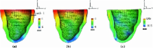
          In this work, we introduce a modified Holzapfel-Ogden hyperelastic constitutive model for ventricular myocardium that accounts for residual stresses, and we investigate the effects of residual stresses in diastole using a magnetic resonance imaging–derived model of the human left ventricle (LV). We adopt an invariant-based constitutive modelling approach and treat the left ventricular myocardium as a non-homogeneous, fibre-reinforced, incompressible material. Because in vivo images provide the configuration of the LV in a loaded state even in diastole, an inverse analysis is used to determine the corresponding unloaded reference configuration. The residual stress in this unloaded state is estimated by two different methods. One is based on three-dimensional strain measurements in a local region of the canine LV, and the other uses the opening angle method for a cylindrical tube. We find that including residual stress in the model changes the stress distributions across the myocardium and that whereas both methods yield qualitatively similar changes, there are quantitative differences between the two approaches. Although the effects of residual stresses are relatively small in diastole, the model can be extended to explore the full impact of residual stress on LV mechanical behaviour for the whole cardiac cycle as more experimental data become available. In addition, although not considered here, residual stresses may also play a larger role in models that account for tissue growth and remodelling.

          Related collections

          Most cited references25

          • Record: found
          • Abstract: found
          • Article: not found

          Single-beat estimation of end-diastolic pressure-volume relationship: a novel method with potential for noninvasive application.

          Whereas end-systolic and end-diastolic pressure-volume relations (ESPVR, EDPVR) characterize left ventricular (LV) pump properties, clinical utility of these relations has been hampered by the need for invasive measurements over a range of pressure and volumes. We propose a single-beat approach to estimate the whole EDPVR from one measured volume-pressure (Vm and Pm) point. Ex vivo EDPVRs were measured from 80 human hearts of different etiologies (normal, congestive heart failure, left ventricular assist device support). Independent of etiology, when EDPVRs were normalized (EDPVRn) by appropriate scaling of LV volumes, EDPVRns were nearly identical and were optimally described by the relation EDP = An.EDV (Bn), with An = 28.2 mmHg and Bn = 2.79. V0 (the volume at the pressure of approximately 0 mmHg) was predicted by using the relation V0 = Vm.(0.6 - 0.006.Pm) and V30 by V30 = V0 + (Vm,n - V0)/(Pm/An) (1/Bn). The entire EDPVR of an individual heart was then predicted by forcing the curve through Vm, Pm, and the predicted V0 and V30. This technique was applied prospectively to the ex vivo human EDPVRs not used in determining optimal An and Bn values and to 36 in vivo human, 12 acute and 14 chronic canine, and 80 in vivo and ex vivo rat studies. The root-mean-square error (RMSE) in pressure between measured and predicted EDPVRs over the range of 0-40 mmHg was < 3 mmHg of measured EDPVR in all settings, indicating a good predictive value of this approach. Volume-normalized EDPVRs have a common shape, despite different etiology and species. This allows the entire curve to be predicted by a new method with a potential for noninvasive application. The results are most accurate when applied to groups of hearts rather than to individual hearts.
            Bookmark
            • Record: found
            • Abstract: found
            • Article: not found

            Shear properties of passive ventricular myocardium.

            We examined the shear properties of passive ventricular myocardium in six pig hearts. Samples (3 x 3 x 3 mm) were cut from adjacent regions of the lateral left ventricular midwall, with sides aligned with the principal material axes. Four cycles of sinusoidal simple shear (maximum shear displacements of 0.1-0.5) were applied separately to each specimen in two orthogonal directions. Resulting forces along the three axes were measured. Three specimens from each heart were tested in different orientations to cover all six modes of simple shear deformation. Passive myocardium has nonlinear viscoelastic shear properties with reproducible, directionally dependent softening as strain is increased. Shear properties were clearly anisotropic with respect to the three principal material directions: passive ventricular myocardium is least resistant to simple shear displacements imposed in the plane of the myocardial layers and most resistant to shear deformations that produce extension of the myocyte axis. Comparison of results for the six different shear modes suggests that simple shear deformation is resisted by elastic elements aligned with the microstructural axes of the tissue.
              Bookmark
              • Record: found
              • Abstract: found
              • Article: not found

              Extracellular matrix of the human aortic media: an ultrastructural histochemical and immunohistochemical study of the adult aortic media.

              Aortic distensability is the key to normal aortic function and relates to the lamellar unit in the media. However, the organization of the extracellular matrix components in these lamellar units, which are largely responsible for the distensability, is insufficiently known, especially in the human. We therefore performed a detailed ultrastructural analysis of these components. Thoracic aortas of 56 individuals (age 45-74 years), none of whom suffered from aortic disease, were studied by immunoelectron microscopy of elastin, collagen types I, III, IV, V, and VI, fibronectin, and fibrillin-1, and by ultrastructural histochemistry of proteoglycans, which were further characterized by enzymatic digestion. The elastic lamellae were closely associated with thick collagen fibers containing types I, III, and V collagen. Between these collagen fibers, numerous complex, circumferentially oriented streaks of elastin protruded from the lamellae. In contrast to what is usually reported in the aortas of experimental animals, the smooth muscle cells preferentially adhered to these ill-defined streaks rather than directly to the solid lamellae. Fibrillin-1- and type VI collagen-containing bundles of microfibrils (oxytalan fibers) were also involved in the smooth muscle cell-elastin contact. The smooth muscle cells were invested by basal lamina-like layers connecting them to each other as well as to the oxytalan fibers. Unexpectedly, these layers were abundantly labeled by anti-fibronectin, whereas type IV collagen, a specific basement membrane component, was mainly found in larger, flocculent deposits. The proteoglycans present were collagen-associated dermatan sulfate proteoglycan, cell-associated heparan sulfate proteoglycan, and interstitial chondroitin sulfate proteoglycan. Our observations demonstrate that the extracellular matrix in the human aorta is extremely complex and therefore differs from most descriptions based on experimental animals. They serve as reference for future studies on aortic diseases, such as aneurysmas and dissections. Copyright 2000 Wiley-Liss, Inc.
                Bookmark

                Author and article information

                Contributors
                xiaoyu.luo@glasgow.ac.uk
                Journal
                Biomech Model Mechanobiol
                Biomech Model Mechanobiol
                Biomechanics and Modeling in Mechanobiology
                Springer Berlin Heidelberg (Berlin/Heidelberg )
                1617-7959
                1617-7940
                23 April 2013
                23 April 2013
                2014
                : 13
                : 99-113
                Affiliations
                [ ]School of  Civil Engineering, Xinjiang University, Xinjiang, China
                [ ]School of Mathematics and Statistics, University of Glasgow, Glasgow, UK
                [ ]Leon H. Charney Division of Cardiology, Department of Medicine, New York University School of Medicine, New York, NY USA
                [ ]Institute of Cardiovascular and Medical Sciences, University of Glasgow, Glasgow, UK
                [ ]SV Lab, School of Aerospace Engineering, Xi’an Jiaotong University, Xi’an, China
                Article
                488
                10.1007/s10237-013-0488-x
                3880672
                23609894
                8fcd5773-03bf-4f07-9097-3a00c0b610be
                © The Author(s) 2013

                Open AccessThis article is distributed under the terms of the Creative Commons Attribution License which permits any use, distribution, and reproduction in any medium, provided the original author(s) and the source are credited.

                History
                : 17 December 2012
                : 27 March 2013
                Categories
                Original Paper
                Custom metadata
                © Springer-Verlag Berlin Heidelberg 2014

                Biophysics
                residual stress,finite strain ,finite stress,left ventricle
                Biophysics
                residual stress, finite strain , finite stress, left ventricle

                Comments

                Comment on this article