1
views
0
recommends
+1 Recommend
0 collections
    0
    shares
      • Record: found
      • Abstract: found
      • Article: found
      Is Open Access

      Leukoaraiosis Is Not Associated With Recovery From Aphasia in the First Year After Stroke

      research-article

      Read this article at

      Bookmark
          There is no author summary for this article yet. Authors can add summaries to their articles on ScienceOpen to make them more accessible to a non-specialist audience.

          Abstract

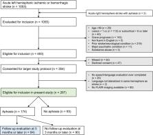
          After a stroke, individuals with aphasia often recover to a certain extent over time. This recovery process may be dependent on the health of surviving brain regions. Leukoaraiosis (white matter hyperintensities on MRI reflecting cerebral small vessel disease) is one indication of compromised brain health and is associated with cognitive and motor impairment. Previous studies have suggested that leukoaraiosis may be a clinically relevant predictor of aphasia outcomes and recovery, although findings have been inconsistent. We investigated the relationship between leukoaraiosis and aphasia in the first year after stroke. We recruited 267 patients with acute left hemispheric stroke and coincident fluid attenuated inversion recovery MRI. Patients were evaluated for aphasia within 5 days of stroke, and 174 patients presented with aphasia acutely. Of these, 84 patients were evaluated at ∼3 months post-stroke or later to assess longer-term speech and language outcomes. Multivariable regression models were fit to the data to identify any relationships between leukoaraiosis and initial aphasia severity, extent of recovery, or longer-term aphasia severity. We found that leukoaraiosis was present to varying degrees in 90% of patients. However, leukoaraiosis did not predict initial aphasia severity, aphasia recovery, or longer-term aphasia severity. The lack of any relationship between leukoaraiosis severity and aphasia recovery may reflect the anatomical distribution of cerebral small vessel disease, which is largely medial to the white matter pathways that are critical for speech and language function.

          Related collections

          Most cited references42

          • Record: found
          • Abstract: not found
          • Article: not found

          Guidelines, criteria, and rules of thumb for evaluating normed and standardized assessment instruments in psychology.

            Bookmark
            • Record: found
            • Abstract: found
            • Article: not found

            MR signal abnormalities at 1.5 T in Alzheimer's dementia and normal aging.

            The type, frequency, and extent of MR signal abnormalities in Alzheimer's disease and normal aging are a subject of controversy. With a 1.5-MR unit we studied 12 Alzheimer patients, four subjects suffering from multiinfarct dementia and nine age-matched controls. Punctate or early confluent high-signal abnormalities in the deep white matter, noted in 60% of both Alzheimer patients and controls, were unrelated to the presence of hypertension or other vascular risk factors. A significant number of Alzheimer patients exhibited a more extensive smooth "halo" of periventricular hyperintensity when compared with controls (p = .024). Widespread deep white-matter hyperintensity (two patients) and extensive, irregular periventricular hyperintensity (three patients) were seen in multiinfarct dementia. Areas of high signal intensity affecting hippocampal and sylvian cortex were also present in five Alzheimer and two multiinfarct dementia patients, but absent in controls. Discrete, small foci of deep white-matter hyperintensity are not characteristic of Alzheimer's disease nor do they appear to imply a vascular cause for the dementing illness. The frequently observed "halo" of periventricular hyperintensity in Alzheimer's disease may be of diagnostic importance. High-signal abnormalities in specific cortical regions are likely to reflect disease processes localized to those structures.
              Bookmark
              • Record: found
              • Abstract: found
              • Article: not found

              Cerebral small vessel disease: from pathogenesis and clinical characteristics to therapeutic challenges.

              The term cerebral small vessel disease refers to a group of pathological processes with various aetiologies that affect the small arteries, arterioles, venules, and capillaries of the brain. Age-related and hypertension-related small vessel diseases and cerebral amyloid angiopathy are the most common forms. The consequences of small vessel disease on the brain parenchyma are mainly lesions located in the subcortical structures such as lacunar infarcts, white matter lesions, large haemorrhages, and microbleeds. Because lacunar infarcts and white matter lesions are easily detected by neuroimaging, whereas small vessels are not, the term small vessel disease is frequently used to describe the parenchyma lesions rather than the underlying small vessel alterations. This classification, however, restricts the definition of small vessel disease to ischaemic lesions and might be misleading. Small vessel disease has an important role in cerebrovascular disease and is a leading cause of cognitive decline and functional loss in the elderly. Small vessel disease should be a main target for preventive and treatment strategies, but all types of presentation and complications should be taken into account. Copyright 2010 Elsevier Ltd. All rights reserved.
                Bookmark

                Author and article information

                Contributors
                Role: Role: Role: Role: Role: Role:
                Role: Role:
                Role: Role:
                Role: Role:
                Role: Role:
                Role: Role:
                Role: Role:
                Role: Role: Role:
                Role: Role: Role: Role: Role: Role: Role:
                Journal
                Neurobiol Lang (Camb)
                Neurobiol Lang (Camb)
                nol
                Neurobiology of Language
                MIT Press (One Broadway, 12th Floor, Cambridge, Massachusetts 02142, USA journals-info@mit.edu )
                2641-4368
                2023
                31 October 2023
                : 4
                : 4
                : 536-549
                Affiliations
                [1]School of Medicine, Vanderbilt University, Nashville, TN, USA
                [2]Department of Hearing and Speech Sciences, Vanderbilt University Medical Center, Nashville, TN, USA
                [3]Department of Radiology, Vanderbilt University Medical Center, Nashville, TN, USA
                [4]School of Health and Rehabilitation Sciences, University of Queensland, Brisbane, Australia
                Author notes

                Competing Interests: The authors have declared that no competing interests exist.

                * Corresponding Author: smwilson@ 123456uq.edu.au

                Handling Editor: Julius Fridriksson

                Author information
                https://orcid.org/0000-0002-1389-2525
                https://orcid.org/0000-0001-9447-4431
                https://orcid.org/0000-0001-8822-0005
                https://orcid.org/0000-0002-6178-6356
                https://orcid.org/0000-0003-3854-5121
                https://orcid.org/0000-0003-2939-3875
                https://orcid.org/0000-0001-9884-2852
                Article
                nol_a_00115
                10.1162/nol_a_00115
                10631799
                8ff12071-20ef-4615-a2e0-f0ccaf736be2
                © 2023 Massachusetts Institute of Technology

                This is an open-access article distributed under the terms of the Creative Commons Attribution 4.0 International License, which permits unrestricted use, distribution, and reproduction in any medium, provided the original work is properly cited. For a full description of the license, please visit https://creativecommons.org/licenses/by/4.0/.

                History
                : 14 October 2022
                : 28 June 2023
                Page count
                Pages: 14
                Funding
                Funded by: National Institute on Deafness and Other Communication Disorders, DOI 10.13039/100000055;
                Award ID: R01 DC013270
                Award Recipient :
                Categories
                Research Article
                Custom metadata
                Brito, A. C., Levy, D. F., Schneck, S. M., Entrup, J. L., Onuscheck, C. F., Casilio, M., de Riesthal, M., Davis, L. T., & Wilson, S. M. (2023). Leukoaraiosis is not associated with recovery from aphasia in the first year after stroke. Neurobiology of Language, 4(4), 536–549. https://doi.org/10.1162/nol_a_00115

                aphasia,brain health,cerebral small vessel disease,leukoaraiosis,stroke

                Comments

                Comment on this article