12
views
0
recommends
+1 Recommend
0 collections
    0
    shares
      • Record: found
      • Abstract: found
      • Article: found
      Is Open Access

      Structural Integrity of the Greek Key Motif in βγ-Crystallins Is Vital for Central Eye Lens Transparency

      research-article

      Read this article at

      Bookmark
          There is no author summary for this article yet. Authors can add summaries to their articles on ScienceOpen to make them more accessible to a non-specialist audience.

          Abstract

          Background

          We highlight an unrecognized physiological role for the Greek key motif, an evolutionarily conserved super-secondary structural topology of the βγ-crystallins. These proteins constitute the bulk of the human eye lens, packed at very high concentrations in a compact, globular, short-range order, generating transparency. Congenital cataract (affecting 400,000 newborns yearly worldwide), associated with 54 mutations in βγ-crystallins, occurs in two major phenotypes nuclear cataract, which blocks the central visual axis, hampering the development of the growing eye and demanding earliest intervention, and the milder peripheral progressive cataract where surgery can wait. In order to understand this phenotypic dichotomy at the molecular level, we have studied the structural and aggregation features of representative mutations.

          Methods

          Wild type and several representative mutant proteins were cloned, expressed and purified and their secondary and tertiary structural details, as well as structural stability, were compared in solution, using spectroscopy. Their tendencies to aggregate in vitro and in cellulo were also compared. In addition, we analyzed their structural differences by molecular modeling in silico.

          Results

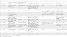
          Based on their properties, mutants are seen to fall into two classes. Mutants A36P, L45PL54P, R140X, and G165fs display lowered solubility and structural stability, expose several buried residues to the surface, aggregate in vitro and in cellulo, and disturb/distort the Greek key motif. And they are associated with nuclear cataract. In contrast, mutants P24T and R77S, associated with peripheral cataract, behave quite similar to the wild type molecule, and do not affect the Greek key topology.

          Conclusion

          When a mutation distorts even one of the four Greek key motifs, the protein readily self-aggregates and precipitates, consistent with the phenotype of nuclear cataract, while mutations not affecting the motif display ‘native state aggregation’, leading to peripheral cataract, thus offering a protein structural rationale for the cataract phenotypic dichotomy “distort motif, lose central vision”.

          Related collections

          Most cited references50

          • Record: found
          • Abstract: found
          • Article: found
          Is Open Access

          Cat-Map: putting cataract on the map

          Lens opacities, or cataract(s), may be inherited as a classic Mendelian disorder usually with early-onset or, more commonly, acquired with age as a multi-factorial or complex trait. Many genetic forms of cataract have been described in mice and other animal models. Considerable progress has been made in mapping and identifying the genes and mutations responsible for inherited forms of cataract, and genetic determinants of age-related cataract are beginning to be discovered. To provide a convenient and accurate summary of current information focused on the increasing genetic complexity of Mendelian and age-related cataract we have created an online chromosome map and reference database for cataract in humans and mice (Cat-Map).
            Bookmark
            • Record: found
            • Abstract: found
            • Article: not found

            Genetics of crystallins: cataract and beyond.

            The crystallins were discovered more than 100 years ago by Mörner (1893. Untersuchungen der Proteinsubstanzen in den lichtbrechenden Medien des Auges. Z. Physiol. Chem. 18, 61-106) as the main structural proteins of the vertebrate eye lens. Since that time the major mammalian crystallins referred to as alpha-, beta-, and gamma-crystallins were characterized with respect to their genetic organization, regulation of their expression pattern and participation in several diseases. In recent years, more and more crystallins have also been identified outside the lens. Evolutionary analysis has demonstrated the relationship of crystallins to proteins involved in protection against stress. The alpha-crystallins form large complexes up to 1Mio Da; they are built up by two subunits referred to as alphaA- and alphaB-crystallins. These subunits are encoded by individual genes, Cryaa and Cryab being localized on different chromosomes and members of the small heat-shock protein family. The alphaA-crystallin is considered to be a molecular chaperone. It is expressed mainly in the lens - mutations in the Cryaa gene lead to recessive or dominant cataracts. In contrast, the alphaB-crystallin is rather ubiquitously expressed; mutations in the Cryab gene are associated with a broad variety of neurological, cardiac and muscular disorders. The beta/gamma-crystallin super family is encoded by at least 14 genes; the proteins are characterized by four Greek key motifs. In mammals, these genes are not only organized as individual genes (Cryba1, Cryba2, Crygf, Crygs, CrygN), but also in duplets (Cryba4-Crybb1 and Crybb2-Crybb3) and in one major cluster (Cryga-Cryge). The various Cryb and Cryg genes are considered to have been evolved by various duplications of the Greek key encoding units. The two main families are distinguished by the fact that each Greek key motif in the Cryb genes is encoded by one exon, whereas two motifs are encoded by one single exon in the Cryg genes. An intermediate between these subfamilies is CrygN encoding the first two Greek key motifs by individual exons, but the others by one single exon. Mutations in the Cryb/Cryg genes lead mainly to an opacification of the eye lens. In some Cryg mutants evidence was presented that the formation of large amyloid-like intranuclear inclusions containing the altered gamma-crystallins is a key event in cataract formation. Cataract formation, caused by Cryg mutations is further characterized by stopping the secondary lens fiber differentiation as indicated by the presence of remnants of cell nuclei, which are usually degraded in secondary fiber cells. Moreover, additional clinical features are being increasingly reported since these crystallins are found outside the eye: the betaB2-crystallin (previously referred to the basic principle crystallin) is also involved in neurogenesis and male infertility. For some of the beta/gamma-crystallins, Ca(2+)-binding properties have been discussed; however, it is an unsolved question whether these crystallins serve as Ca(2+) stores in vivo. Enzyme crystallins are enzymes, which have been recruited to the lens and are expressed there in high concentrations. The mu- and zeta-crystallins (gene symbols: Crym and Cryz, respectively) are discussed as examples for mammals. Mutations in the human CRYM gene lead to non-syndromic deafness, and mutations in the Cryz gene of guinea pigs cause cataracts.
              Bookmark
              • Record: found
              • Abstract: not found
              • Article: not found

              Genetic origins of cataract.

                Bookmark

                Author and article information

                Contributors
                Role: Editor
                Journal
                PLoS One
                PLoS ONE
                plos
                plosone
                PLoS ONE
                Public Library of Science (San Francisco, USA )
                1932-6203
                2013
                6 August 2013
                : 8
                : 8
                : e70336
                Affiliations
                [1 ]Hyderabad Eye Research Foundation, L. V. Prasad Eye Institute, Hyderabad, India
                [2 ]Molecular Biophysics Unit, Indian Institute of Science, Bangalore, India
                [3 ]Novarus Discoveries Pvt Ltd, Hyderabad, India
                [4 ]University of Miami Miller School of Medicine, Miami, Florida, United States of America
                University of Arkansas for Medical Sciences, United States of America
                Author notes

                Competing Interests: Dr SC is employed by Novarus Discoveries Pvt Ltd. This does not alter the authors’ adherence to all the PLOS ONE policies on sharing data and materials.

                Conceived and designed the experiments: DB NS VPRV. Performed the experiments: VPRV GA SC. Analyzed the data: DB NS VPRV GA SC VT. Contributed reagents/materials/analysis tools: NS SC. Wrote the paper: DB VPRV NS.

                Article
                PONE-D-13-17271
                10.1371/journal.pone.0070336
                3735602
                23936409
                904490b1-2ceb-485e-beff-d2d2d1015c99
                Copyright @ 2013

                This is an open-access article distributed under the terms of the Creative Commons Attribution License, which permits unrestricted use, distribution, and reproduction in any medium, provided the original author and source are credited.

                History
                : 29 April 2013
                : 13 June 2013
                Page count
                Pages: 10
                Funding
                This work was supported by grants from the Department of Biotechnology, India, and the Hyderabad Eye Research Foundation. The funders had no role in study design, data collection and analysis, decision to publish, or preparation of the manuscript.
                Categories
                Research Article
                Biology
                Biochemistry
                Proteins
                Globular Proteins
                Biophysics
                Protein Folding
                Genetics
                Genetic Mutation
                Genetics of Disease
                Medicine
                Diagnostic Medicine
                Pathology
                General Pathology
                Molecular Pathology
                Ophthalmology
                Cataracts and Other Lens Disorders

                Uncategorized
                Uncategorized

                Comments

                Comment on this article