45
views
0
recommends
+1 Recommend
0 collections
    0
    shares
      • Record: found
      • Abstract: found
      • Article: found
      Is Open Access

      High Cholesterol Deteriorates Bone Health: New Insights into Molecular Mechanisms

      review-article

      Read this article at

      Bookmark
          There is no author summary for this article yet. Authors can add summaries to their articles on ScienceOpen to make them more accessible to a non-specialist audience.

          Abstract

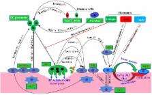
          Many epidemiological studies show a positive connection between cardiovascular diseases and risk of osteoporosis, suggesting a role of hyperlipidemia and/or hypercholesterolemia in regulating osteoporosis. The majority of the studies indicated a correlation between high cholesterol and high LDL-cholesterol level with low bone mineral density, a strong predictor of osteoporosis. Similarly, bone metastasis is a serious complication of cancer for patients. Several epidemiological and basic studies have established that high cholesterol is associated with increased cancer risk. Moreover, osteoporotic bone environment predisposes the cancer cells for metastatic growth in the bone microenvironment. This review focuses on how cholesterol and cholesterol-lowering drugs (statins) regulate the functions of bone residential osteoblast and osteoclast cells to augment or to prevent bone deterioration. Moreover, this study provides an insight into molecular mechanisms of cholesterol-mediated bone deterioration. It also proposes a potential mechanism by which cellular cholesterol boosts cancer-induced bone metastasis.

          Related collections

          Most cited references85

          • Record: found
          • Abstract: found
          • Article: not found

          Leptin inhibits bone formation through a hypothalamic relay: a central control of bone mass.

          Gonadal failure induces bone loss while obesity prevents it. This raises the possibility that bone mass, body weight, and gonadal function are regulated by common pathways. To test this hypothesis, we studied leptin-deficient and leptin receptor-deficient mice that are obese and hypogonadic. Both mutant mice have an increased bone formation leading to high bone mass despite hypogonadism and hypercortisolism. This phenotype is dominant, independent of the presence of fat, and specific for the absence of leptin signaling. There is no leptin signaling in osteoblasts but intracerebroventricular infusion of leptin causes bone loss in leptin-deficient and wild-type mice. This study identifies leptin as a potent inhibitor of bone formation acting through the central nervous system and therefore describes the central nature of bone mass control and its disorders.
            Bookmark
            • Record: found
            • Abstract: found
            • Article: not found

            Relationship between osteoporosis and cardiovascular disease in postmenopausal women.

            In the placebo group of the MORE study, including 2576 postmenopausal women (mean age, 66.5 years), the authors describe a strong linear association between the severity grade of osteoporosis (from low BMD to presence of severe vertebral fractures) and the future risk of cardiovascular events. Accordingly, treatment of postmenopausal osteoporosis should include consideration of measures to prevent adverse cardiovascular outcomes. Observations indicate an inverse association between BMD and the severity of peripheral atherosclerosis in postmenopausal women. The predictive value of osteoporosis and its different severity stages for the risk of acute cardiovascular events remains unknown. Participants were 2576 women (mean age, 66.5 years) assigned to placebo and followed for 4 years in an osteoporosis treatment trial. Those with at least one vertebral fracture or total hip BMD T score < or = -2.5 at baseline were defined as having osteoporosis, whereas those without vertebral fracture and total hip BMD T score between -2.5 and -1 were defined as having low bone mass. The primary outcome for these posthoc analyses was the incidence of adjudicated fatal or nonfatal cardiovascular events. After adjustment for potential confounders, women with osteoporosis had a 3.9-fold (95% CI, 2.0-7.7; p < 0.001) increased risk for cardiovascular events compared with women with low bone mass. Under the same boundaries, a total hip BMD T score < or = -2.5 versus a T score between -2.5 and -1 was associated with a 2.1-fold (95% CI, 1.2-3.6; p < 0.01) increase in risk, whereas presence of at least one vertebral fracture versus no vertebral fracture at baseline was associated with a 3.0-fold (95% CI, 1.8-5.1; p < 0.001) increase in risk. The risk of cardiovascular events increased incrementally with the number and increasing severity of baseline vertebral fractures (both p < 0.001). Postmenopausal women with osteoporosis are at an increased risk for cardiovascular events that is proportional to the severity of osteoporosis at the time of the diagnosis. Treatment of postmenopausal osteoporosis should include consideration of measures to prevent cardiovascular outcomes.
              Bookmark
              • Record: found
              • Abstract: found
              • Article: not found

              Matrix vesicles and calcification.

              Matrix vesicles (MVs) are extracellular, 100 nM in diameter, membrane-invested particles selectively located at sites of initial calcification in cartilage, bone, and predentin. The first crystals of apatitic bone mineral are formed within MVs close to the inner surfaces of their investing membranes. Matrix vesicle biogenesis occurs by polarized budding and pinching-off of vesicles from specific regions of the outer plasma membranes of differentiating growth plate chondrocytes, osteoblasts, and odontoblasts. Polarized release of MVs into selected areas of developing matrix determines the nonrandom distribution of calcification. Initiation of the first mineral crystals, within MVs (phase 1), is augmented by the activity of MV phosphatases (eg, alkaline phosphatase, adenosine triphosphatase and pyrophosphatase) plus calcium-binding molecules (eg, annexin I and phosphatidyl serine), all of which are concentrated in or near the MV membrane. Phase 2 of biologic mineralization begins with crystal release through the MV membrane, exposing preformed hydroxyapatite crystals to the extracellular fluid. The extracellular fluid normally contains sufficient Ca2+ and PO4(3-) to support continuous crystal proliferation, with preformed crystals serving as nuclei (templates) for the formation of new crystals by a process of homologous nucleation. In diseases such as osteoarthritis, crystal deposition arthritis, and atherosclerosis, MVs initiate pathologic calcification, which, in turn, augments disease progression.
                Bookmark

                Author and article information

                Contributors
                URI : http://frontiersin.org/people/u/197120
                Journal
                Front Endocrinol (Lausanne)
                Front Endocrinol (Lausanne)
                Front. Endocrinol.
                Frontiers in Endocrinology
                Frontiers Media S.A.
                1664-2392
                23 October 2015
                2015
                : 6
                : 165
                Affiliations
                [1] 1Department of Biochemistry, School of Life Sciences, Central University of Rajasthan , Rajasthan, India
                Author notes

                Edited by: Jakob Starup-Linde, Aarhus Universitetshospital, Denmark

                Reviewed by: Gudrun Stenbeck, Brunel University, UK; Archana Sanjay, University of Connecticut Health Center, USA

                *Correspondence: Chandi C. Mandal, chandicmandal@ 123456gmail.com

                Specialty section: This article was submitted to Bone Research, a section of the journal Frontiers in Endocrinology

                Article
                10.3389/fendo.2015.00165
                4617053
                26557105
                90f9fb3e-e529-4bd8-9903-9d45ce6c9db4
                Copyright © 2015 Mandal.

                This is an open-access article distributed under the terms of the Creative Commons Attribution License (CC BY). The use, distribution or reproduction in other forums is permitted, provided the original author(s) or licensor are credited and that the original publication in this journal is cited, in accordance with accepted academic practice. No use, distribution or reproduction is permitted which does not comply with these terms.

                History
                : 29 May 2015
                : 08 October 2015
                Page count
                Figures: 3, Tables: 3, Equations: 0, References: 105, Pages: 11, Words: 7804
                Funding
                Funded by: University Grants Commission 10.13039/501100001501
                Award ID: 30-49/2014 (BSR)
                Funded by: Department of Biotechnology, Ministry of Science and Technology 10.13039/501100001407
                Award ID: 6242 P9/RGCB/PMD/DBT/CCML/2015
                Categories
                Endocrinology
                Review

                Endocrinology & Diabetes
                osteoporosis,bone mineral density,bone remodeling,bone metastasis,osteoblast,osteoclast,cholesterol,statins

                Comments

                Comment on this article