2
views
0
recommends
+1 Recommend
0 collections
    0
    shares
      • Record: found
      • Abstract: found
      • Article: found
      Is Open Access

      Acute myocardial infarction not attributed to coronary artery disease: A seldom initial presentation of a left ventricular myxoma

      case-report

      Read this article at

      Bookmark
          There is no author summary for this article yet. Authors can add summaries to their articles on ScienceOpen to make them more accessible to a non-specialist audience.

          Abstract

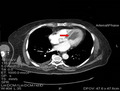
          Although myxoma represents the most frequent non‐malignant cardiac primary tumor; it is extremely rare met in the left ventricle. Clinical features of the neoplasm extend from symptomless to critical signs of either ischemia or embolism. We describe here an unusual case of a huge left ventricular myxoma in a 68‐year‐old man, presented with clinical and ECG findings of an inferior wall myocardial infarction. The patient was primarily referred to our institution for coronary angiography, which showed no coronary artery disease. Further examinations revealed a left ventricular mass as the possible source of embolization, thus the patient underwent surgery for tumor excision. The postoperative course was unremarkable. A bibliographical analysis demonstrated that those tumors are rare but treatable causes of embolic myocardial infarction, thus profound clinical intuition, proper utilization of imaging modalities, administration of anticoagulants preoperatively, as well immediate surgical removal are justified.

          Abstract

          Myocardial infarction due to left ventricular myxoma. High clinical suspicion to establish the diagnosis is warranted.

          Related collections

          Most cited references18

          • Record: found
          • Abstract: found
          • Article: not found

          The complex of myxomas, spotty pigmentation, and endocrine overactivity.

          Of 40 patients (16 males and 24 females), 29 had cardiac myxoma(s), 14 had skin pigmentation (lentigo and several types of nevi) which also commonly affected the lips, 6 had skin myxoma(s), and 12 had both pigmentation and myxoma(s); 18 had primary pigmented nodular adrenocortical disease (Cushing syndrome was present in 11); 10 had myxoid mammary fibroadenomas; 9 had testicular tumor(s) (large-cell calcifying Sertoli cell tumor, Leydig cell tumor, or adrenocortical rest tumor, or a combination); and 4 had pituitary adenoma with gigantism or acromegaly. The maximum number of conditions present together was five, occurring in two patients; each of the remaining patients had at least two of the conditions. The overlap, in this sizeable number of patients, of various combinations of the same rare or very rare conditions unlikely to occur together by chance with any degree of frequency is striking evidence for a unique syndrome. The patients were young (mean age at diagnosis of the first component, 18 years). Pathologic involvement tended to be multicentric (heart and skin) and bilateral in paired organs (adrenal, breast, and testis). Thirteen patients (32%) are alive and well. Twelve patients are alive but with complications of cardiac myxoma (in 8), testicular tumors (in 2), residual Cushing syndrome (in 1), or bilateral pulmonary nodules (in 1). Twelve patients are dead: 9 of cardiac myxoma, 1 of intracranial (nonpituitary) tumor, and 2 postoperatively. The status of three is unknown.
            Bookmark
            • Record: found
            • Abstract: not found
            • Article: not found

            Cardiac tumours: diagnosis and management.

              Bookmark
              • Record: found
              • Abstract: found
              • Article: not found

              Cardiac tumors: optimal cardiac MR sequences and spectrum of imaging appearances.

              This article reviews the optimal cardiac MRI sequences for and the spectrum of imaging appearances of cardiac tumors. Recent technologic advances in cardiac MRI have resulted in the rapid acquisition of images of the heart with high spatial and temporal resolution and excellent myocardial tissue characterization. Cardiac MRI provides optimal assessment of the location, functional characteristics, and soft-tissue features of cardiac tumors, allowing accurate differentiation of benign and malignant lesions.
                Bookmark

                Author and article information

                Contributors
                spiliopoulos@med.uth.gr
                Journal
                Clin Case Rep
                Clin Case Rep
                10.1002/(ISSN)2050-0904
                CCR3
                Clinical Case Reports
                John Wiley and Sons Inc. (Hoboken )
                2050-0904
                04 May 2021
                May 2021
                : 9
                : 5 ( doiID: 10.1002/ccr3.v9.5 )
                : e04029
                Affiliations
                [ 1 ] Department of Thoracic and Cardiovascular Surgery Faculty of Medicine School of Health Sciences University of Thessaly Larissa Greece
                [ 2 ] Department of Cardiology Faculty of Medicine School of Health Sciences University of Thessaly Larissa Greece
                [ 3 ] Department of Surgery Faculty of Medicine School of Health Sciences University of Thessaly Larissa Greece
                Author notes
                [*] [* ] Correspondence

                Kyriakos Spiliopoulos, University of Thessaly, School of Medicine, Department of Thoracic and Cardiovacsular Surgery, Panepistimiou 3 (Viopolis), Post Box 1400, GR‐41500, Larissa, Greece.

                Email: spiliopoulos@ 123456med.uth.gr

                Author information
                https://orcid.org/0000-0003-4332-258X
                Article
                CCR34029
                10.1002/ccr3.4029
                8142312
                34084484
                9443e61e-ca7f-4071-bf65-d607e0b0347a
                © 2021 The Authors. Clinical Case Reports published by John Wiley & Sons Ltd.

                This is an open access article under the terms of the http://creativecommons.org/licenses/by/4.0/ License, which permits use, distribution and reproduction in any medium, provided the original work is properly cited.

                History
                : 22 January 2021
                : 03 August 2020
                : 18 February 2021
                Page count
                Figures: 4, Tables: 0, Pages: 5, Words: 3057
                Categories
                Case Report
                Case Reports
                Custom metadata
                2.0
                May 2021
                Converter:WILEY_ML3GV2_TO_JATSPMC version:6.0.2 mode:remove_FC converted:24.05.2021

                cardiac tumors,myocardial infarction,myxoma,surgical excision

                Comments

                Comment on this article