61
views
0
recommends
+1 Recommend
0 collections
    0
    shares
      • Record: found
      • Abstract: found
      • Article: found
      Is Open Access

      Optical Coherence Tomography Angiography Vessel Density in Healthy, Glaucoma Suspect, and Glaucoma Eyes

      research-article

      Read this article at

      Bookmark
          There is no author summary for this article yet. Authors can add summaries to their articles on ScienceOpen to make them more accessible to a non-specialist audience.

          Abstract

          Purpose

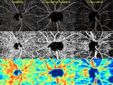
          The purpose of this study was to compare retinal nerve fiber layer (RNFL) thickness and optical coherence tomography angiography (OCT-A) retinal vasculature measurements in healthy, glaucoma suspect, and glaucoma patients.

          Methods

          Two hundred sixty-one eyes of 164 healthy, glaucoma suspect, and open-angle glaucoma (OAG) participants from the Diagnostic Innovations in Glaucoma Study with good quality OCT-A images were included. Retinal vasculature information was summarized as a vessel density map and as vessel density (%), which is the proportion of flowing vessel area over the total area evaluated. Two vessel density measurements extracted from the RNFL were analyzed: (1) circumpapillary vessel density (cpVD) measured in a 750-μm-wide elliptical annulus around the disc and (2) whole image vessel density (wiVD) measured over the entire image. Areas under the receiver operating characteristic curves (AUROC) were used to evaluate diagnostic accuracy.

          Results

          Age-adjusted mean vessel density was significantly lower in OAG eyes compared with glaucoma suspects and healthy eyes. (cpVD: 55.1 ± 7%, 60.3 ± 5%, and 64.2 ± 3%, respectively; P < 0.001; and wiVD: 46.2 ± 6%, 51.3 ± 5%, and 56.6 ± 3%, respectively; P < 0.001). For differentiating between glaucoma and healthy eyes, the age-adjusted AUROC was highest for wiVD (0.94), followed by RNFL thickness (0.92) and cpVD (0.83). The AUROCs for differentiating between healthy and glaucoma suspect eyes were highest for wiVD (0.70), followed by cpVD (0.65) and RNFL thickness (0.65).

          Conclusions

          Optical coherence tomography angiography vessel density had similar diagnostic accuracy to RNFL thickness measurements for differentiating between healthy and glaucoma eyes. These results suggest that OCT-A measurements reflect damage to tissues relevant to the pathophysiology of OAG.

          Related collections

          Most cited references50

          • Record: found
          • Abstract: found
          • Article: found
          Is Open Access

          Split-spectrum amplitude-decorrelation angiography with optical coherence tomography

          Amplitude decorrelation measurement is sensitive to transverse flow and immune to phase noise in comparison to Doppler and other phase-based approaches. However, the high axial resolution of OCT makes it very sensitive to the pulsatile bulk motion noise in the axial direction. To overcome this limitation, we developed split-spectrum amplitude-decorrelation angiography (SSADA) to improve the signal-to-noise ratio (SNR) of flow detection. The full OCT spectrum was split into several narrower bands. Inter-B-scan decorrelation was computed using the spectral bands separately and then averaged. The SSADA algorithm was tested on in vivo images of the human macula and optic nerve head. It significantly improved both SNR for flow detection and connectivity of microvascular network when compared to other amplitude-decorrelation algorithms.
            Bookmark
            • Record: found
            • Abstract: found
            • Article: not found

            Optical coherence tomography angiography of optic disc perfusion in glaucoma.

            To compare optic disc perfusion between normal subjects and subjects with glaucoma using optical coherence tomography (OCT) angiography and to detect optic disc perfusion changes in glaucoma.
              Bookmark
              • Record: found
              • Abstract: found
              • Article: not found

              Optical Coherence Tomography Angiography of the Peripapillary Retina in Glaucoma.

              Vascular factors may have important roles in the pathophysiology of glaucoma. A practical method for the clinical evaluation of ocular perfusion is needed to improve glaucoma management.
                Bookmark

                Author and article information

                Journal
                Invest Ophthalmol Vis Sci
                Invest. Ophthalmol. Vis. Sci
                iovs
                iovs
                iovs
                Investigative Ophthalmology & Visual Science
                The Association for Research in Vision and Ophthalmology
                0146-0404
                1552-5783
                13 July 2016
                July 2016
                : 57
                : 9
                : OCT451-OCT459
                Affiliations
                [1 ]Hamilton Glaucoma Center Shiley Eye Institute, Department of Ophthalmology, University of California, San Diego, La Jolla, California, United States
                [2 ]Department of Ophthalmology and Otorhinolaryngology, Federal University of Minas Gerais, Belo Horizonte, Brazil
                [3 ]Department of Ophthalmology, Haeundae Paik Hospital, Inje University College of Medicine, Busan, South Korea
                [4 ]Casey Eye Institute, Oregon Health & Science University, Portland, Oregon, United States
                Author notes
                Correspondence: Robert N. Weinreb, Hamilton Glaucoma Center, Department of Ophthalmology, University of California, San Diego, 9500 Gilman Drive, La Jolla, CA, 92093-0946, USA; rweinreb@ 123456ucsd.edu .
                AY and LMZ are joint first authors.

                AY and LMZ are joint first authors.

                Article
                iovs-57-101-33 IOVS-15-18944
                10.1167/iovs.15-18944
                4968912
                27409505
                963ea420-2eda-4a57-ae59-684a10fed7dd

                This work is licensed under a Creative Commons Attribution-NonCommercial-NoDerivatives 4.0 International License.

                History
                : 14 December 2015
                : 29 March 2016
                Categories
                Articles

                glaucoma,oct angiography,rnfl
                glaucoma, oct angiography, rnfl

                Comments

                Comment on this article