14
views
0
recommends
+1 Recommend
0 collections
    0
    shares
      • Record: found
      • Abstract: found
      • Article: found
      Is Open Access

      Cell–Cell Interaction Proteins (Gap Junctions, Tight Junctions, and Desmosomes) and Water Transporter Aquaporin 4 in Meningothelial Cells of the Human Optic Nerve

      research-article

      Read this article at

      Bookmark
          There is no author summary for this article yet. Authors can add summaries to their articles on ScienceOpen to make them more accessible to a non-specialist audience.

          Abstract

          Purpose

          Meningothelial cells (MECs) play a central role in the maintenance of cerebrospinal fluid (CSF) homeostasis and in physiological and pathophysiological processes within the subarachnoid space (SAS) linking them to optic nerve (ON) pathologies. Still, not much is known about their structural properties that might enable MECs to perform specific functions within the ON microenvironment.

          Methods

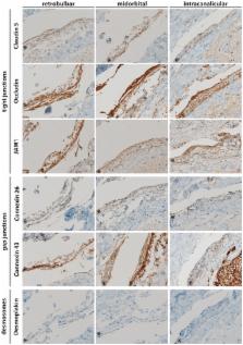
          For closer characterization of the structural properties of the human MEC layer in the arachnoid, we performed immunohistological analyses to evaluate the presence of cell–cell interaction markers, namely, markers for tight junctions (JAM1, Occludin, and Claudin 5), gap junctions (Connexin 26 and 43), and desmosomes (Desmoplakin) as well as for water channel marker aquaporin 4 (AQP4) in retrobulbar, midorbital, and intracanalicular human ON sections.

          Results

          MECs displayed immunopositivity for markers of tight junctions (JAM1, Occludin, and Claudin 5) and gap junctions (Connexin 26 and 43) as well as for AQP4 water channels. However, no immunopositivity was found for Desmoplakin.

          Conclusion

          MECs are connected via tight junctions and gap junctions, and they possess AQP4 water channels. The presence of these proteins emphasizes the important function of MECs within the ON microenvironment as part of the meningeal barrier. Beyond this barrier function, the expression of these proteins by MECs supports a broader role of these cells in signal transduction and CSF clearance pathways within the ON microenvironment.

          Related collections

          Most cited references16

          • Record: found
          • Abstract: found
          • Article: not found

          Endothelial cell-to-cell junctions: molecular organization and role in vascular homeostasis.

          Intercellular junctions mediate adhesion and communication between adjoining endothelial and epithelial cells. In the endothelium, junctional complexes comprise tight junctions, adherens junctions, and gap junctions. The expression and organization of these complexes depend on the type of vessels and the permeability requirements of perfused organs. Gap junctions are communication structures, which allow the passage of small molecular weight solutes between neighboring cells. Tight junctions serve the major functional purpose of providing a "barrier" and a "fence" within the membrane, by regulating paracellular permeability and maintaining cell polarity. Adherens junctions play an important role in contact inhibition of endothelial cell growth, paracellular permeability to circulating leukocytes and solutes. In addition, they are required for a correct organization of new vessels in angiogenesis. Extensive research in the past decade has identified several molecular components of the tight and adherens junctions, including integral membrane and intracellular proteins. These proteins interact both among themselves and with other molecules. Here, we review the individual molecules of junctions and their complex network of interactions. We also emphasize how the molecular architectures and interactions may represent a mechanistic basis for the function and regulation of junctions, focusing on junction assembly and permeability regulation. Finally, we analyze in vivo studies and highlight information that specifically relates to the role of junctions in vascular endothelial cells.
            Bookmark
            • Record: found
            • Abstract: found
            • Article: not found

            Architecture of arachnoid trabeculae, pillars, and septa in the subarachnoid space of the human optic nerve: anatomy and clinical considerations.

            To describe the anatomy and the arrangement of the arachnoid trabeculae, pillars, and septa in the subarachnoid space of the human optic nerve and to consider their possible clinical relevance for cerebrospinal fluid dynamics and fluid pressure in the subarachnoid space of the human optic nerve. Postmortem study with a total of 12 optic nerves harvested from nine subjects without ocular disease. All optic nerves used in this study were obtained no later than 7 hours after death, following qualified consent for necropsy. The study was performed with transmission (TEM) and scanning electron microscopy (SEM). The subarachnoid space of the human optic nerve contains a variety of trabeculae, septa, and stout pillars that are arranged between the arachnoid and the pia layers of the meninges of the nerve. They display a considerable numeric and structural variability depending on their location within the different portions of the optic nerve. In the bulbar segment (ampulla), adjacent to the globe, a dense and highly ramified meshwork of delicate trabeculae is arranged in a reticular fashion. Between the arachnoid trabeculae, interconnecting velum-like processes are observed. In the mid-orbital segment of the orbital portion, the subarachnoid space is subdivided, and can appear even loosely chambered by broad trabeculae and velum-like septa at some locations. In the intracanalicular segment additionally, few stout pillars and single round trabeculae are observed. The subarachnoid space of the human optic nerve is not a homogeneous and anatomically empty chamber filled with cerebrospinal fluid, but it contains a complex system of arachnoid trabeculae and septa that divide the subarachnoid space. The trabeculae, septa, and pillars, as well as their arrangement described in this study, may have a role in the cerebrospinal fluid dynamics between the subarachnoid space of the optic nerve and the chiasmal cistern and may contribute to the understanding of the pathophysiology of asymmetric and unilateral papilloedema. All the structures described are of such delicate character that they can not even be visualised with high resolution magnetic resonance imaging (MRI).
              Bookmark
              • Record: found
              • Abstract: found
              • Article: not found

              Junctions in the meninges and marginal glia.

              The meninges of various mammals were prepared for examination with the electronmicroscope by thin sectioning or freeze-fracturing. Particular attention was given to the distribution of tight junctions in order to determine the basis for the meningeal barrier between the blood circulating in dural vessels and the cerebrospinal fluid in the subarachnoid space. While some dural blood vessels are fenestrated, those in the subarachnoid space are not and their component endothelial cells are joined by an extensive system of tight junctions. An extensive and continuous system of tight junctions was also found in a layer of specialized cells at the border of the arachnoid with the dura. This arachnoid barrier layer is apparently the only basis of the meningeal barrier because often cellular layers in the dura and arachnoid lack tight junctions although they are linked by gap junctions and desmosomes. In particular, tight junctions are lacking at the border of the "subdural space" which is actually a fascial plane within the dura. Tight junctions are also lacking between astrocytes at the surface of the brain but these cells are linked by gap junctions and a new type of intercellular junction. The distribution of these junctions, as well as assemblies of intramembranous particles at the astrocytic border, raises the question whether this layer might have a role in the exchange of certain substances between the brain and cerebrospinal fluid.
                Bookmark

                Author and article information

                Contributors
                Journal
                Front Neurol
                Front Neurol
                Front. Neurol.
                Frontiers in Neurology
                Frontiers Media S.A.
                1664-2295
                29 June 2017
                2017
                : 8
                : 308
                Affiliations
                [1] 1Department of Ophthalmology, Kantonsspital Aarau , Aarau, Switzerland
                [2] 2Department of Biomedicine, Ocular Pharmacology and Physiology, University Hospital Basel , Basel, Switzerland
                [3] 3Department of Ophthalmology, University Hospital Basel , Basel, Switzerland
                Author notes

                Edited by: Kenneth Shindler, University of Pennsylvania, United States

                Reviewed by: Jayasri Das Sarma, Indian Institute of Science Education and Research-Kolkata, India; Gregory Wu, Washington University in St. Louis, United States

                *Correspondence: Hanspeter E. Killer, hanspeter.killer@ 123456ksa.ch ; Peter Meyer, peter.meyer@ 123456usb.ch

                These authors have contributed equally to this work.

                Specialty section: This article was submitted to Neuro-Ophthalmology, a section of the journal Frontiers in Neurology

                Article
                10.3389/fneur.2017.00308
                5489558
                28706505
                98cc740c-497b-4390-9b13-9e6138ad0209
                Copyright © 2017 Zeleny, Kohler, Neutzner, Killer and Meyer.

                This is an open-access article distributed under the terms of the Creative Commons Attribution License (CC BY). The use, distribution or reproduction in other forums is permitted, provided the original author(s) or licensor are credited and that the original publication in this journal is cited, in accordance with accepted academic practice. No use, distribution or reproduction is permitted which does not comply with these terms.

                History
                : 09 March 2017
                : 13 June 2017
                Page count
                Figures: 5, Tables: 0, Equations: 0, References: 20, Pages: 9, Words: 3551
                Categories
                Neuroscience
                Original Research

                Neurology
                optic nerve degeneration,optic nerve microenvironment,meninges,meningothelial cells,cell–cell interactions,aquaporin 4,cerebrospinal fluid homeostasis

                Comments

                Comment on this article