30
views
0
recommends
+1 Recommend
0 collections
    0
    shares
      • Record: found
      • Abstract: found
      • Article: found
      Is Open Access

      Effect of ST36 Acupuncture on Hyperventilation-Induced CO 2 Reactivity of the Basilar and Middle Cerebral Arteries and Heart Rate Variability in Normal Subjects

      research-article

      Read this article at

      Bookmark
          There is no author summary for this article yet. Authors can add summaries to their articles on ScienceOpen to make them more accessible to a non-specialist audience.

          Abstract

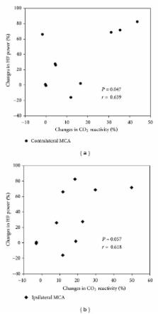
          This study was conducted to verify the effect of acupuncture on cerebral haemodynamics to provide evidence for the use of acupuncture treatment as a complementary therapy for the high-risk stroke population. The effect of ST36 acupuncture treatment on the hyperventilation-induced CO 2 reactivity of the basilar and middle cerebral arteries was studied in 10 healthy male volunteers (mean age, 25.2 ± 1.5 years) using a transcranial Doppler sonography with an interval of 1 week between measurements, and a portable ECG monitoring system was used to obtain ECG data simultaneously. The CO 2 reactivity of the basilar and middle cerebral arteries increased significantly after ST36 acupuncture treatment, whereas the mean arterial blood pressure and pulse rate did not change significantly. The high-frequency power significantly increased after ST36 acupuncture treatment, and the percentage increase of high-frequency power correlated significantly with the percentage increase in the CO 2 reactivity of the contralateral middle cerebral artery. These data suggest that ST36 acupuncture treatment increases CO 2 reactivity, indicating improvement of vasodilatory potential of the cerebral vasculature to compensate for fluctuations caused by changes in external conditions. The increase in parasympathetic tone by ST36 acupuncture treatment is responsible for this therapeutic effect.

          Related collections

          Most cited references45

          • Record: found
          • Abstract: found
          • Article: found

          Cerebrovascular Reactivity in Degenerative and Vascular Dementia: A Transcranial Doppler Study

          Background: An impairment of cerebral microvessels is reported both in normal ageing and in senescence-associated processes, as well as in Alzheimer’s disease (AD) and vascular dementia (VaD). The aim of this study was to explore cerebral hemodynamics by transcranial Doppler in VaD and AD, compared with age-matched control subjects. Methods: Transcranial Doppler was investigated in all patients in the basal condition. Cerebral vasoreactivity to hyper- and hypocapnia was evaluated with CO 2 mixture inhalation followed by hyperventilation. Results: We studied 60 AD and 58 VaD patients and 62 nondemented controls. Both AD and VaD subjects showed lower flow velocities (FV) and higher pulsatility indices (PI) as compared with controls. Lower total vasomotor reactivity and lower response to hypercapnia were observed in the AD and VaD groups as compared with controls. AD and VaD patients did not show significant differences in FV, PI values or cerebral vasoreactivity. Conclusions: Reduced FV and increased PI with a significant vasoreactivity reduction in VaD and AD patients are indicators of impairment of cerebral microvasculature circulation in both diseases. The identification of vascular function impairment in all kinds of dementia could be of help in identifying patients who would thus benefit more from specific therapeutic approaches.
            Bookmark
            • Record: found
            • Abstract: found
            • Article: not found

            Dependency of blood flow velocity in the middle cerebral artery on end-tidal carbon dioxide partial pressure--a transcranial ultrasound Doppler study.

            The end-tidal carbon dioxide partial pressure (PCO2) response curves for the flow velocity in the middle cerebral artery were studied in 31 normal subjects with transcranial Doppler techniques. An exponential curve with an exponent of 0.034 mm Hg-1 was found to be a good fit to the recorded data. By means of this relationship, recordings of flow velocity in cerebral arteries can be normalized to a standard value of PCO2. Physiological aspects of cerebrovascular reactivity to PCO2 and the clinical implications of the PCO2 response curve are discussed. The normal material provides a reference for assessing pathological responses.
              Bookmark
              • Record: found
              • Abstract: found
              • Article: not found

              Cerebral vasomotor reactivity and cerebral white matter lesions in the elderly.

              The pathogenesis of white matter lesions is still uncertain, but an ischemic-hypoxic cause has been suggested. Cerebral vasomotor reactivity reflects the compensatory dilatory mechanism of the intracerebral arterioles to a vasodilatory stimulus and provides a more sensitive hemodynamic index than the level of resting flow. The authors determined the association between vasomotor reactivity and white matter lesions in 73 consecutive individuals from the Rotterdam Scan Study who also participated in the Rotterdam Study, a large population-based prospective follow-up study of individuals > or =55 years old. Vasomotor reactivity was measured by means of CO2-enhanced transcranial Doppler, and in all individuals axial T1*-, T2*-, and proton density (PD)-weighted MRI scans (1.5 T) were obtained. White matter lesions were scored according to location, size, and number by two independent readers. Vasomotor reactivity was inversely associated with the deep subcortical and total periventricular white matter lesions (OR 0.5, 95% CI 0.3 to 1.1; and OR 0.7, 95% CI 0.4 to 1.1, respectively). A strong association was found between impaired vasomotor reactivity and periventricular white matter lesions adjacent to the lateral ventricular wall (OR 0.6, 95% CI 0.4 to 1.0; p = 0.001). No association was found with periventricular white matter lesions near the frontal and occipital horns. Our data confirm the association between vasomotor reactivity and white matter lesions and support the hypothesis that some white matter lesions may be associated with hemodynamic ischemic injury to the brain.
                Bookmark

                Author and article information

                Journal
                Evid Based Complement Alternat Med
                Evid Based Complement Alternat Med
                ECAM
                Evidence-based Complementary and Alternative Medicine : eCAM
                Hindawi Publishing Corporation
                1741-427X
                1741-4288
                2014
                14 July 2014
                14 July 2014
                : 2014
                : 574986
                Affiliations
                Department of Cardiology and Neurology of Korean Medicine, College of Korean Medicine, Kyung Hee University, No. 26 Kyungheedae-ro, Dongdaemun-gu, Seoul 130-872, Republic of Korea
                Author notes
                *Sang-Kwan Moon: skmoon@ 123456khu.ac.kr

                Academic Editor: Andreas Sandner-Kiesling

                Author information
                http://orcid.org/0000-0002-4404-4158
                http://orcid.org/0000-0002-1680-3524
                http://orcid.org/0000-0002-8611-568X
                http://orcid.org/0000-0003-3149-9759
                Article
                10.1155/2014/574986
                4123599
                25132861
                99232ec2-c823-4c20-8fdf-d8a0c829458d
                Copyright © 2014 Sang-Ho Hyun et al.

                This is an open access article distributed under the Creative Commons Attribution License, which permits unrestricted use, distribution, and reproduction in any medium, provided the original work is properly cited.

                History
                : 26 May 2014
                : 25 June 2014
                Categories
                Research Article

                Complementary & Alternative medicine
                Complementary & Alternative medicine

                Comments

                Comment on this article