2
views
0
recommends
+1 Recommend
0 collections
    0
    shares
      • Record: found
      • Abstract: found
      • Article: found
      Is Open Access

      Epicardial macroreentrant atrial tachycardia involving a large left atrial roof diverticulum: insights using high-resolution coherent mapping

      research-article

      Read this article at

      Bookmark
          There is no author summary for this article yet. Authors can add summaries to their articles on ScienceOpen to make them more accessible to a non-specialist audience.

          Abstract

          A 75-year-old woman with hypertrophic cardiomyopathy and symptomatic atrial tachycardia (AT) was admitted for first-time catheter ablation (Supplementary material online, Figure S1 ). Entrainment during AT-1 (cycle length: CL = 230 ms) from both the left atrial (LA) roof and mitral annulus (MA) revealed that both sites were within the circuit. A high-resolution electroanatomic map of the left atrium was created using a PentaRay catheter with the CARTO mapping system (Biosense Webster, Diamond Bar, CA, USA). Activation mapping was performed, and the Coherent module (CARTO, Biosense Webster, Diamond Bar, CA, USA) was used to display the colour and conduction velocity vectors for the electrical wave propagation. 1 Video 1 shows the Coherent propagation map during AT-1, which was consistent with a dual loop macroreentry with roof-dependent and MA circuits. After creation of a linear ablation lesion set from the MA to the anterior left superior pulmonary vein, AT-1 transformed to another AT (AT-2, Supplementary material online, Figure S2 ) with a different propagation pattern. Activation mapping of the endocardial LA clearly demonstrated that the activation started from the summit of a large diverticulum on the LA roof ( Figure 1 , 2 , Supplementary material online, Figure S3 ), propagating to its base and then the left atrium and right atrium (Video 2). The mapped propagation time in the left atrium was 235 ms, which did not cover the total AT-2 CL (265 ms), and the 30 ms difference was regarded as the conduction time through the epicardial tissue. Entrainment mapping demonstrated that the circuit of AT-2 included the proximal and distal coronary sinus and diverticulum (Supplementary material online, Figures S4 and S5). Therefore, like AT-1, AT-2 was speculated to also be a counter clockwise perimitral atrial flutter. Since the endocardial aspect of the left atrium was blocked, propagation was felt to use epicardial connections along the anterior LA wall utilizing the diverticulum before entering the endocardial left atrium. AT-2 was terminated during radiofrequency catheter ablation around the base of diverticulum, after which AT-2 was rendered non-inducible. Voltage mapping demonstrated large areas of bipolar voltage abnormality at the anterior and perimitral LA (Supplementary material online, Figure S6 ), and which may have provided the substrate for sustaining tachycardia. High-density Coherent mapping was able to visualize the circuit of an unusual macroreentrant AT utilizing an epicardial connection associated with an LA roof diverticulum, which to the best of our knowledge, has not been previously reported.
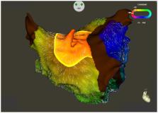
 Figure 1 Computed tomography of the LA and a large diverticulum (anterior–posterior view). LA, left atrium; LAA, left atrial appendage; LSPV, left superior pulmonary vein; RIPV, right inferior pulmonary vein; RSPV, right superior pulmonary vein.
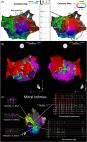
 Figure 2 Coherent propagation map during AT-2. Supplementary material Supplementary material is available at European Heart Journal - Case Reports online. Supplementary Material ytaa268_Supplementary_Data Click here for additional data file.

          Related collections

          Most cited references1

          • Record: found
          • Abstract: found
          • Article: found
          Is Open Access

          Identification of critical isthmus using coherent mapping in patients with scar‐related atrial tachycardia

          Abstract Introduction Accurate identification of slow conducting regions in patients with scar‐related atrial tachycardia (AT) is difficult using conventional electrogram annotation for cardiac electroanatomic mapping (EAM). Estimating delays between neighboring mapping sites is a potential option for activation map computation. We describe our initial experience with CARTO 3 Coherent Mapping (Biosense Webster Inc,) in the ablation of complex ATs. Methods Twenty patients (58 ± 10 y/o, 15 males) with complex ATs were included. We created three‐dimensional EAMs using CARTO 3 system with CONFIDENSE and a high‐resolution mapping catheter (Biosense Webster Inc). Local activation time and coherent maps were used to aid in the identification of conduction isthmus (CI) and focal origin sites. System‐defined slow or nonconducting zones and CI, defined by concealed entrainment (postpacing interval < 20 ms), CV < 0.3 m/s and local fractionated electrograms were evaluated. Results Twenty‐six complex ATs were mapped (mean: 1.3 ± 0.7 maps/pt; 4 focal, 22 isthmus‐dependent). Coherent mapping was better in identifying CI/breakout sites where ablation terminated the tachycardia (96.2% vs 69.2%; P = .010) and identified significantly more CI (mean/chamber 2.0 ± 1.1 vs 1.0 ± 0.7; P < .001) with narrower width (19.8 ± 10.5 vs 43.0 ± 23.9 mm; P < .001) than conventional mapping. Ablation at origin and CI sites was successful in 25 (96.2%) with long‐term recurrence in 25%. Conclusions Coherent mapping with conduction velocity vectors derived from adjacent mapping sites significantly improved the identification of CI sites in scar‐related ATs with isthmus‐dependent re‐entry better than conventional mapping. It may be used in conjunction with conventional mapping strategies to facilitate recognition of slow conduction areas and critical sites that are important targets of ablation.
            Bookmark

            Author and article information

            Contributors
            Role: Handling Editor
            Role: Editor
            Journal
            Eur Heart J Case Rep
            Eur Heart J Case Rep
            ehjcr
            European Heart Journal: Case Reports
            Oxford University Press
            2514-2119
            October 2020
            19 September 2020
            19 September 2020
            : 4
            : 5
            : 1-2
            Affiliations
            Arrhythmia Advanced Therapy Center, AOI Universal Hospital , 2-9-1 Tamachi Kawasaki-ku, Kawasaki City, Kanagawa 210-0822, Japan
            Arrhythmia Advanced Therapy Center, AOI Universal Hospital , 2-9-1 Tamachi Kawasaki-ku, Kawasaki City, Kanagawa 210-0822, Japan
            Arrhythmia Advanced Therapy Center, AOI Universal Hospital , 2-9-1 Tamachi Kawasaki-ku, Kawasaki City, Kanagawa 210-0822, Japan
            Arrhythmia Advanced Therapy Center, AOI Universal Hospital , 2-9-1 Tamachi Kawasaki-ku, Kawasaki City, Kanagawa 210-0822, Japan
            Author notes
            Corresponding author. Tel: +81 44 277 5511, Fax: +81 44 277 5568, Email: sin_go_sin_go_@ 123456hotmail.com
            Author information
            http://orcid.org/0000-0001-6964-0653
            http://orcid.org/0000-0002-1023-2708
            http://orcid.org/0000-0002-0136-483X
            Article
            ytaa268
            10.1093/ehjcr/ytaa268
            7780435
            9b02ceb7-06ed-4894-a338-aabf2c4d5be8
            © The Author(s) 2020. Published by Oxford University Press on behalf of the European Society of Cardiology.

            This is an Open Access article distributed under the terms of the Creative Commons Attribution Non-Commercial License ( http://creativecommons.org/licenses/by-nc/4.0/), which permits non-commercial re-use, distribution, and reproduction in any medium, provided the original work is properly cited. For commercial re-use, please contact journals.permissions@oup.com

            History
            : 24 May 2020
            : 16 June 2020
            : 16 July 2020
            Page count
            Pages: 2
            Categories
            Images Cardio
            Arrhythmias / Electrophysiology
            AcademicSubjects/MED00200

            Comments

            Comment on this article