1
views
0
recommends
+1 Recommend
0 collections
    0
    shares
      • Record: found
      • Abstract: found
      • Article: found
      Is Open Access

      Quantification of Endogenous Brain Tissue Displacement Imaging by Radiofrequency Ultrasound

      research-article

      Read this article at

      Bookmark
          There is no author summary for this article yet. Authors can add summaries to their articles on ScienceOpen to make them more accessible to a non-specialist audience.

          Abstract

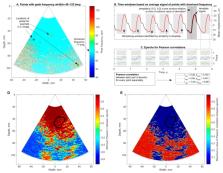
          The purpose of this paper is a quantification of displacement parameters used in the imaging of brain tissue endogenous motion using ultrasonic radiofrequency (RF) signals. In a preclinical study, an ultrasonic diagnostic system with RF output was equipped with dedicated signal processing software and subject head–ultrasonic transducer stabilization. This allowed the use of RF scanning frames for the calculation of micrometer-range displacements, excluding sonographer-induced motions. Analysis of quantitative displacement estimates in dynamical phantom experiments showed that displacements of 55 µm down to 2 µm were quantified as confident according to Pearson correlation between signal fragments (minimum p ≤ 0.001). The same algorithm and scanning hardware were used in experiments and clinical imaging which allows translating phantom results to Alzheimer’s disease patients and healthy elderly subjects as examples. The confident quantitative displacement waveforms of six in vivo heart-cycle episodes ranged from 8 µm up to 263 µm (Pearson correlation p ≤ 0.01). Displacement time sequences showed promising possibilities to evaluate the morphology of endogenous displacement signals at each point of the scanning plane, while displacement maps—regional distribution of displacement parameters—were essential for tissue characterization.

          Related collections

          Most cited references16

          • Record: found
          • Abstract: found
          • Article: not found
          Is Open Access

          WFUMB guidelines and recommendations for clinical use of ultrasound elastography: Part 1: basic principles and terminology.

          Conventional diagnostic ultrasound images of the anatomy (as opposed to blood flow) reveal differences in the acoustic properties of soft tissues (mainly echogenicity but also, to some extent, attenuation), whereas ultrasound-based elasticity images are able to reveal the differences in the elastic properties of soft tissues (e.g., elasticity and viscosity). The benefit of elasticity imaging lies in the fact that many soft tissues can share similar ultrasonic echogenicities but may have different mechanical properties that can be used to clearly visualize normal anatomy and delineate pathologic lesions. Typically, all elasticity measurement and imaging methods introduce a mechanical excitation and monitor the resulting tissue response. Some of the most widely available commercial elasticity imaging methods are 'quasi-static' and use external tissue compression to generate images of the resulting tissue strain (or deformation). In addition, many manufacturers now provide shear wave imaging and measurement methods, which deliver stiffness images based upon the shear wave propagation speed. The goal of this review is to describe the fundamental physics and the associated terminology underlying these technologies. We have included a questions and answers section, an extensive appendix, and a glossary of terms in this manuscript. We have also endeavored to ensure that the terminology and descriptions, although not identical, are broadly compatible across the WFUMB and EFSUMB sets of guidelines on elastography (Bamber et al. 2013; Cosgrove et al. 2013).
            Bookmark
            • Record: found
            • Abstract: found
            • Article: found
            Is Open Access

            The pulsating brain: A review of experimental and clinical studies of intracranial pulsatility

            The maintenance of adequate blood flow to the brain is critical for normal brain function; cerebral blood flow, its regulation and the effect of alteration in this flow with disease have been studied extensively and are very well understood. This flow is not steady, however; the systolic increase in blood pressure over the cardiac cycle causes regular variations in blood flow into and throughout the brain that are synchronous with the heart beat. Because the brain is contained within the fixed skull, these pulsations in flow and pressure are in turn transferred into brain tissue and all of the fluids contained therein including cerebrospinal fluid. While intracranial pulsatility has not been a primary focus of the clinical community, considerable data have accrued over the last sixty years and new applications are emerging to this day. Investigators have found it a useful marker in certain diseases, particularly in hydrocephalus and traumatic brain injury where large changes in intracranial pressure and in the biomechanical properties of the brain can lead to significant changes in pressure and flow pulsatility. In this work, we review the history of intracranial pulsatility beginning with its discovery and early characterization, consider the specific technologies such as transcranial Doppler and phase contrast MRI used to assess various aspects of brain pulsations, and examine the experimental and clinical studies which have used pulsatility to better understand brain function in health and with disease.
              Bookmark
              • Record: found
              • Abstract: found
              • Article: not found

              Methods for estimation of subsample time delays of digitized echo signals.

              Time delay estimation (TDE) is commonly performed in practice by crosscorrelation of digitized echo signals. Since time delays are generally not integral multiples of the sampling period, the location of the largest sample of the crosscorrelation function (ccf) is an inexact estimator of the location of the peak. Therefore, one must interpolate between the samples of the ccf to improve the estimation precision. Using theory and simulations, we review and compare the performance of several methods for interpolation of the ccf. The maximum likelihood approach to interpolation is the application of a reconstruction filter to the discrete ccf. However, this method can only be approximated in practice and can be computationally intensive. For these reasons, a simple method is widely used that involves fitting a parabola (or other curve) to samples of the ccf in the neighborhood of its peak. We describe and compare two curve-fitting methods: parabolic and cosine interpolation. Curve-fitting interpolation can yield biased time-delay estimates, which may preclude the use of these methods in some applications. The artifactual effect of these bias errors on elasticity imaging by elastography is discussed. We demonstrate that reconstructive interpolation is unbiased. An iterative implementation of the reconstruction procedure is proposed that can reduce the computation time significantly.
                Bookmark

                Author and article information

                Journal
                Diagnostics (Basel)
                Diagnostics (Basel)
                diagnostics
                Diagnostics
                MDPI
                2075-4418
                21 January 2020
                February 2020
                : 10
                : 2
                : 57
                Affiliations
                [1 ]Biomedical Engineering Institute, Kaunas University of Technology, K. Baršausko Str. 59-455, LT-51423 Kaunas, Lithuania; monika.makunaite@ 123456ktu.edu (M.M.); m.baranauskas@ 123456ktu.lt (M.B.); arunas.lukosevicius@ 123456ktu.lt (A.L.); sakalauskas.andrius@ 123456yahoo.com (A.S.)
                [2 ]Department of Neurology, Lithuanian University of Health Sciences, A. Mickevičiaus Str. 9, LT-44307 Kaunas, Lithuania; vaidas.matijosaitis@ 123456lsmuni.lt (V.M.); Daiva.Rastenyte@ 123456lsmuni.lt (D.R.)
                Author notes
                Author information
                https://orcid.org/0000-0002-9481-1773
                https://orcid.org/0000-0003-0204-4886
                https://orcid.org/0000-0002-5603-2357
                Article
                diagnostics-10-00057
                10.3390/diagnostics10020057
                7168898
                31973031
                9b33a36b-3732-4aca-a563-36c1db49340a
                © 2020 by the authors.

                Licensee MDPI, Basel, Switzerland. This article is an open access article distributed under the terms and conditions of the Creative Commons Attribution (CC BY) license ( http://creativecommons.org/licenses/by/4.0/).

                History
                : 30 November 2019
                : 16 January 2020
                Categories
                Article

                brain,transcranial sonography,radiofrequency ultrasound,tissue displacements

                Comments

                Comment on this article