10
views
0
recommends
+1 Recommend
0 collections
    0
    shares
      • Record: found
      • Abstract: found
      • Article: found
      Is Open Access

      Gastric heterotopic pancreas and stromal tumors smaller than 3 cm in diameter: clinical and computed tomography findings

      research-article

      Read this article at

      Bookmark
          There is no author summary for this article yet. Authors can add summaries to their articles on ScienceOpen to make them more accessible to a non-specialist audience.

          Abstract

          Background

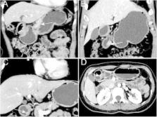
          Identifying gastric heterotopic pancreas and stromal tumors is difficult. Few studies have reported computed tomography (CT) findings for differentiating lesions less than 3 cm in diameter. In this study, we aimed to identify clinical characteristics and CT findings that can differentiate gastric heterotopic pancreatic lesions from stromal tumors less than 3 cm in diameter.

          Methods

          A total of 132 patients with pathologically confirmed gastric heterotopic pancreas ( n = 66) and stromal tumors ( n = 66) were included. Each group was divided into primary ( n = 50) and validation cohort ( n = 16). Clinical characteristics and CT findings were retrospectively reviewed. CT findings included location, border, contour, growth pattern, enhancement pattern and grade, the enhancement value of tumor, enhancement ratio of tumor, and enhancement ratio of tumor to pancreas in venous phase. The findings in the two groups were compared using the Pearson χ 2 test or Student t-test. Receiver operating characteristic curves were used to determine areas under the curve and optimal cut-offs.

          Results

          Significant differences were observed between heterotopic pancreas and stromal tumors in the distribution of tumor location, border, contour (all P < 0.001), enhancement values ( P < 0.001), enhancement ratios of tumors ( P < 0.001), and enhancement ratios of tumors to pancreas ( P < 0.001). No significant differences existed in growth pattern ( P = 0.203). The area under the curve differed significantly between enhancement ratio of tumor to pancreas and enhancement ratio ( P = 0.030). There were significant differences in above characteristics between two groups in validation cohort.

          Conclusions

          Heterotopic pancreas has characteristic CT features differentiating it from stromal tumors.

          Related collections

          Most cited references31

          • Record: found
          • Abstract: found
          • Article: not found

          Heterotopic Pancreas: Histopathologic Features, Imaging Findings, and Complications

          Heterotopic pancreas is a congenital anomaly in which pancreatic tissue is anatomically separate from the main gland. The most common locations of this displacement include the upper gastrointestinal tract-specifically, the stomach, duodenum, and proximal jejunum. Less common sites are the esophagus, ileum, Meckel diverticulum, biliary tree, mesentery, and spleen. Uncomplicated heterotopic pancreas is typically asymptomatic, with the lesion being discovered incidentally during an unrelated surgery, during an imaging examination, or at autopsy. The most common computed tomographic appearance of heterotopic pancreas is that of a small oval intramural mass with microlobulated margins and an endoluminal growth pattern. The attenuation and enhancement characteristics of these lesions parallel their histologic composition. Acinus-dominant lesions demonstrate avid homogeneous enhancement after intravenous contrast material administration, whereas duct-dominant lesions are hypovascular and heterogeneous. At magnetic resonance imaging, the heterotopic pancreas is isointense to the orthotopic pancreas, with characteristic T1 hyperintensity and early avid enhancement after intravenous gadolinium-based contrast material administration. Heterotopic pancreatic tissue has a rudimentary ductal system in which an orifice is sometimes visible at imaging as a central umbilication of the lesion. Complications of heterotopic pancreas include pancreatitis, pseudocyst formation, malignant degeneration, gastrointestinal bleeding, bowel obstruction, and intussusception. Certain complications may be erroneously diagnosed as malignancy. Paraduodenal pancreatitis is thought to be due to cystic degeneration of heterotopic pancreatic tissue in the medial wall of the duodenum. Recognizing the characteristic imaging features of heterotopic pancreas aids in differentiating it from cancer and thus in avoiding unnecessary surgery. © RSNA, 2017.
            Bookmark
            • Record: found
            • Abstract: found
            • Article: not found

            Ectopic pancreas: CT findings with emphasis on differentiation from small gastrointestinal stromal tumor and leiomyoma.

            To describe the computed tomographic (CT) findings of ectopic pancreas and to identify the features that differentiate it from other similarly manifesting gastric submucosal tumors such as gastrointestinal stromal tumor (GIST) and leiomyoma, which are the most common gastrointestinal submucosal tumors. This retrospective study was approved by the institutional review board and did not require informed consent. CT images of pathologically proved ectopic pancreases (n = 14), GISTs (n = 33), and leiomyomas (n = 7) in the stomach and duodenum were retrospectively reviewed. Analysis of the CT findings included evaluation of the location, contour, growth pattern, border, enhancement pattern, and enhancement grade of the tumor, as well as the presence of surface dimpling, prominent enhancement of overlying mucosa, and low intralesional attenuation. The attenuation of each lesion, the long diameter (LD), the short diameter (SD), and the LD/SD ratio were measured. Among these findings, statistically significant variables were determined by using the chi(2) test (to compare the categoric variables), the Student t test (for quantitative analysis), and the receiver operating characteristic curve (to determine the optimal cutoff of the LD/SD ratio). The typical location (prepyloric antrum and duodenum), endoluminal growth pattern, ill-defined border, prominent enhancement of overlying mucosa, and an LD/SD ratio of greater than 1.4 were found to be significant for differentiating ectopic pancreas from other tumors (P < .05 for each finding). When at least two of these five criteria were used in combination, the sensitivity and specificity for diagnosing ectopic pancreas were 100% (14 of 14) and 82.5% (33 of 40), respectively. When four of these criteria were used, a sensitivity of 42.9% and a specificity of 100% were achieved. By using specific CT criteria, ectopic pancreas can be differentiated from small GIST or leiomyoma with a high degree of accuracy. (c) RSNA, 2009.
              Bookmark
              • Record: found
              • Abstract: found
              • Article: not found

              Heterotopic pancreas in the stomach: a case report and literature review.

              Ectopic pancreas is defined as pancreatic tissue found outside the usual anatomic location of the pancreas. It is often an incidental finding and can be found at different sites in the gastrointestinal tract. It may become clinically evident when complicated by pathologic changes such as inflammation, bleeding, obstruction, and malignant transformation. In this report, a 40 years old woman with epigastric pain due to ectopic pancreatic tissue in the stomach is described. The difficulty of making an accurate diagnosis is highlighted. The patient has remained free of symptoms since she underwent wedge resection of the lesion three years ago. Frozen sections may help in deciding the extent of resection intraoperatively. Although ectopic pancreas is rare, it should be considered in the differential diagnosis of a submucosal gastric tumour.
                Bookmark

                Author and article information

                Contributors
                perfect1989a@163.com
                fengleiyu91@163.com
                41253050@qq.com
                liangpan.1@163.com
                lijingqingqing@163.com
                gaojianbo_cancer@163.com
                Journal
                Cancer Imaging
                Cancer Imaging
                Cancer Imaging
                BioMed Central (London )
                1740-5025
                1470-7330
                7 August 2018
                7 August 2018
                2018
                : 18
                : 26
                Affiliations
                [1 ]GRID grid.412633.1, Department of Radiology, , The First Affiliated Hospital of Zhengzhou University, ; Zhengzhou, 450052 Henan Province China
                [2 ]GRID grid.412633.1, Department of Internal Medicine, , The First Affiliated Hospital of Zhengzhou University, ; Zhengzhou, 450052 Henan Province China
                Article
                161
                10.1186/s40644-018-0161-9
                6081935
                30086800
                9b74f41c-2642-4d51-9165-1eabbe7a18d2
                © The Author(s). 2018

                Open AccessThis article is distributed under the terms of the Creative Commons Attribution 4.0 International License ( http://creativecommons.org/licenses/by/4.0/), which permits unrestricted use, distribution, and reproduction in any medium, provided you give appropriate credit to the original author(s) and the source, provide a link to the Creative Commons license, and indicate if changes were made. The Creative Commons Public Domain Dedication waiver ( http://creativecommons.org/publicdomain/zero/1.0/) applies to the data made available in this article, unless otherwise stated.

                History
                : 12 April 2018
                : 29 July 2018
                Funding
                Funded by: the National Natural Science Foundation of China
                Award ID: No.81701687
                Award Recipient :
                Categories
                Research Article
                Custom metadata
                © The Author(s) 2018

                gastric,heterotopic pancreas,stromal tumor,computed tomography

                Comments

                Comment on this article