38
views
0
recommends
+1 Recommend
0 collections
    0
    shares
      • Record: found
      • Abstract: found
      • Article: found
      Is Open Access

      Alpha oscillation neurofeedback modulates amygdala complex connectivity and arousal in posttraumatic stress disorder

      research-article

      Read this article at

      Bookmark
          There is no author summary for this article yet. Authors can add summaries to their articles on ScienceOpen to make them more accessible to a non-specialist audience.

          Abstract

          Objective

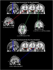
          Electroencephalogram (EEG) neurofeedback aimed at reducing the amplitude of the alpha-rhythm has been shown to alter neural networks associated with posttraumatic stress disorder (PTSD), leading to symptom alleviation. Critically, the amygdala is thought to be one of the central brain regions mediating PTSD symptoms. In the current study, we compare directly patterns of amygdala complex connectivity using fMRI, before and after EEG neurofeedback, in order to observe subcortical mechanisms associated with behavioural and alpha oscillatory changes among patients.

          Method

          We examined basolateral (BLA), centromedial (CMA), and superficial (SFA) amygdala complex resting-state functional connectivity using a seed-based approach via SPM Anatomy Toolbox. Amygdala complex connectivity was measured in twenty-one individuals with PTSD, before and after a 30-minute session of EEG neurofeedback targeting alpha desynchronization.

          Results

          EEG neurofeedback was associated with a shift in amygdala complex connectivity from areas implicated in defensive, emotional, and fear processing/memory retrieval (left BLA and left SFA to the periaqueductal gray, and left SFA to the left hippocampus) to prefrontal areas implicated in emotion regulation/modulation (right CMA to the medial prefrontal cortex). This shift in amygdala complex connectivity was associated with reduced arousal, greater resting alpha synchronization, and was negatively correlated to PTSD symptom severity.

          Conclusion

          These findings have significant implications for developing targeted non-invasive treatment interventions for PTSD patients that utilize alpha oscillatory neurofeedback, showing evidence of neuronal reconfiguration between areas highly implicated in the disorder, in addition to acute symptom alleviation.

          Highlights

          • Alpha desynchronizing neurofeedback was associated with a shift in amygdala complex connectivity.

          • Connectivity shifted from areas implicated in defensive fear processing/memory retrieval, to prefrontal emotion regulation areas.

          • Shift in amygdala complex connectivity was associated with reduced arousal and PTSD symptom severity, and greater resting alpha synchronization after neurofeedback.

          • These findings have significant implications for developing targeted non-invasive treatment interventions for PTSD patients.

          Related collections

          Most cited references71

          • Record: found
          • Abstract: found
          • Article: not found

          Emotional processing in anterior cingulate and medial prefrontal cortex.

          Negative emotional stimuli activate a broad network of brain regions, including the medial prefrontal (mPFC) and anterior cingulate (ACC) cortices. An early influential view dichotomized these regions into dorsal-caudal cognitive and ventral-rostral affective subdivisions. In this review, we examine a wealth of recent research on negative emotions in animals and humans, using the example of fear or anxiety, and conclude that, contrary to the traditional dichotomy, both subdivisions make key contributions to emotional processing. Specifically, dorsal-caudal regions of the ACC and mPFC are involved in appraisal and expression of negative emotion, whereas ventral-rostral portions of the ACC and mPFC have a regulatory role with respect to limbic regions involved in generating emotional responses. Moreover, this new framework is broadly consistent with emerging data on other negative and positive emotions. Published by Elsevier Ltd.
            Bookmark
            • Record: found
            • Abstract: found
            • Article: not found

            Electrophysiological signatures of resting state networks in the human brain.

            Functional neuroimaging and electrophysiological studies have documented a dynamic baseline of intrinsic (not stimulus- or task-evoked) brain activity during resting wakefulness. This baseline is characterized by slow (<0.1 Hz) fluctuations of functional imaging signals that are topographically organized in discrete brain networks, and by much faster (1-80 Hz) electrical oscillations. To investigate the relationship between hemodynamic and electrical oscillations, we have adopted a completely data-driven approach that combines information from simultaneous electroencephalography (EEG) and functional magnetic resonance imaging (fMRI). Using independent component analysis on the fMRI data, we identified six widely distributed resting state networks. The blood oxygenation level-dependent signal fluctuations associated with each network were correlated with the EEG power variations of delta, theta, alpha, beta, and gamma rhythms. Each functional network was characterized by a specific electrophysiological signature that involved the combination of different brain rhythms. Moreover, the joint EEG/fMRI analysis afforded a finer physiological fractionation of brain networks in the resting human brain. This result supports for the first time in humans the coalescence of several brain rhythms within large-scale brain networks as suggested by biophysical studies.
              Bookmark
              • Record: found
              • Abstract: found
              • Article: not found

              Subcortical and cortical brain activity during the feeling of self-generated emotions.

              In a series of [15O]PET experiments aimed at investigating the neural basis of emotion and feeling, 41 normal subjects recalled and re-experienced personal life episodes marked by sadness, happiness, anger or fear. We tested the hypothesis that the process of feeling emotions requires the participation of brain regions, such as the somatosensory cortices and the upper brainstem nuclei, that are involved in the mapping and/or regulation of internal organism states. Such areas were indeed engaged, underscoring the close relationship between emotion and homeostasis. The findings also lend support to the idea that the subjective process of feeling emotions is partly grounded in dynamic neural maps, which represent several aspects of the organism's continuously changing internal state.
                Bookmark

                Author and article information

                Contributors
                Journal
                Neuroimage Clin
                Neuroimage Clin
                NeuroImage : Clinical
                Elsevier
                2213-1582
                14 July 2016
                2016
                14 July 2016
                : 12
                : 506-516
                Affiliations
                [a ]Department of Neuroscience, Western University, London, ON, Canada
                [b ]Department of Psychology, Western University, London, ON, Canada
                [c ]Department of Psychiatry, Western University, London, ON, Canada
                [d ]Department of Medical Imaging, Western University, London, ON, Canada
                [e ]Department of Medial Biophysics, Western University, London, ON, Canada
                [f ]Laboratory of Neurology and Imaging of Cognition, Department of Neuroscience, University of Geneva, Geneva, Switzerland
                [g ]Imaging, Lawson Health Research Institute, London, ON, Canada
                [h ]Department of Diagnostic Imaging, St. Joseph's Healthcare, London, ON, Canada
                [i ]Department of Psychosomatic Medicine and Psychotherapy, Central Institute of Mental Health, Medical Faculty Mannheim Heidelberg University, Mannheim, Germany
                [j ]Canadian Forces, Health Services, Ottawa, Ontario, Canada
                Author notes
                [* ]Corresponding author at: 339 Windermere Road, PO Box 5339, University Hospital, London, ON N6A 5A5, Canada.University Hospital339 Windermere RoadPO Box 5339LondonONN6A 5A5Canada anicho58@ 123456uwo.ca
                Article
                S2213-1582(16)30126-7
                10.1016/j.nicl.2016.07.006
                5030332
                27672554
                9ca88341-ddb9-4c88-b247-c68b9c2d4cfe
                © 2016 The Authors

                This is an open access article under the CC BY-NC-ND license (http://creativecommons.org/licenses/by-nc-nd/4.0/).

                History
                : 30 October 2015
                : 13 April 2016
                : 12 July 2016
                Categories
                Regular Article

                neurofeedback,electroencephalogram,functional mri,functional connectivity,posttraumatic stress disorder,amygdala

                Comments

                Comment on this article