7
views
0
recommends
+1 Recommend
1 collections
    0
    shares
      • Record: found
      • Abstract: found
      • Article: found
      Is Open Access

      A computational modeling method for root canal endoscopy using a specific CBCT filter: A new era in the metaverse of endodontics begins

      research-article
      1 , 2 ,
      Brazilian Dental Journal
      Fundação Odontológica de Ribeirão Preto
      Cone-beam computed tomography, endoscopy, e-Vol DX, metaverse, root canal endoscopy, software

      Read this article at

      ScienceOpenPublisherPMC
      Bookmark
          There is no author summary for this article yet. Authors can add summaries to their articles on ScienceOpen to make them more accessible to a non-specialist audience.

          Abstract

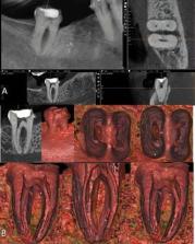
          A contemporary technological revolution has started a new era in the metaverse of Endodontics, a world of virtual operational possibilities that use an exact replica of the natural structures of the maxillofacial complex. This study describes a modeling method for root canal endoscopy using modern cone-beam CT (CBCT) software in a series of clinical cases. The method consists in acquiring thin CBCT slices (0.10mm) in the coronal, sagittal, and axial planes. A specific 3D volume filter, the pulp cavity filter of the e-Vol DX CBCT software, was used to navigate anatomical root canal microstructures, and to scan them using root canal endoscopy. The pulp cavity filter should be set to synchronize CBCT scans from 2D mode - multiplanar reformations (MPR) - to 3D mode - volumetric reconstruction. This filter, when adopting the option of volumetric reconstruction, the developed algorithm leaves the dentin density in transparent mode so that the pulp cavity may be visualized. The algorithm applied performs the suppression (visual) of areas with dentin density. This ensures 3D visualization of the slices and the microanatomy of the root canal, as well as a dynamic navigation throughout the pulp cavity. This computational modeling method adds new resources to Endodontics, which may impact the predictability of root canal treatments positively. The virtual visualization of the internal anatomy of an exact replica of the canal ensures better communications, reliability, and clinical operationalization. Root canal endoscopy using this novel CBCT filter may be used for clinical applications together with innovative digital and virtual-reality resources that will be naturally incorporated into the principles of Endodontics.

          Resumo

          Uma revolução tecnológica contemporânea deu início a uma nova era no metaverso da Endodontia, um mundo de possibilidades operacionais virtuais que utilizam uma réplica exata das estruturas naturais do complexo dentomaxilofacial. Este estudo descreve um método de modelagem computacional para a endoscopia do canal radicular, usando um moderno software de tomografia computadorizada de feixe cônico (TCFC), em uma série de casos clínicos. O método consiste na aquisição de finos slices de TCFC (0,10mm) nos planos coronal, sagital e axial. Um filtro específico de TCFC (filtro cavidade pulpar do software e-Vol DX) foi usado para navegar nas microestruturas anatômicas do canal radicular, e escanear para a aplicação da endoscopia do canal radicular. Este filtro foi configurado para sincronizar as imagens de TCFC em modo 2D - reformações multiplanares (MPR) para o modo 3D - reconstrução volumétrica. O filtro Pulp Cavity ao adotar a opção de reconstrução volumétrica, um algoritmo desenvolvido deixa a densidade dentinária em modo transparente, para que a cavidade pulpar possa ser melhor visualizada. O algoritmo aplicado realiza a supressão (visual) das áreas com densidade dentinária. Este modo de aplicação garante a visualização 3D da microanatomia do canal radicular, bem como permite uma navegação dinâmica por toda a cavidade pulpar. O método de modelagem computacional agrega novos recursos à Endodontia, o que pode impactar positivamente na previsibilidade dos tratamentos endodônticos. A visualização virtual da anatomia interna de uma réplica exata do canal radicular garante melhor comunicação, confiabilidade e operacionalização clínica. O exame de endoscopia do canal radicular com este novo filtro (Pulp cavity) pode ser usada para aplicações clínicas juntamente com recursos digitais e de realidade virtual inovadores que serão naturalmente incorporados aos princípios da Endodontia.

          Related collections

          Most cited references42

          • Record: found
          • Abstract: found
          • Article: not found

          Current challenges and concepts in the preparation of root canal systems: a review.

          Nickel-titanium rotary instruments are important adjuncts in endodontic therapy. This review attempts to identify factors that influence shaping outcomes with these files, such as preoperative root-canal anatomy and instrument tip design. Other, less significant factors include operator experience, rotational speed, and specific instrument sequence. Implications of various working length definitions and desired apical widths are correlated with clinical results. Despite the existence of one ever-present risk factor, dental anatomy, shaping outcomes with nickel-titanium rotary instruments are mostly predictable. Current evidence indicates that wider apical preparations are feasible. Nickel-titanium rotary instruments require a preclinical training period to minimize separation risks and should be used to case-related working lengths and apical widths. However, and despite superior in vitro results, randomized, clinical trials are required to evaluate outcomes when using nickel-titanium instruments.
            Bookmark
            • Record: found
            • Abstract: found
            • Article: not found

            Accuracy of cone beam computed tomography and panoramic and periapical radiography for detection of apical periodontitis.

            The aim of this study was to evaluate the accuracy of imaging methods for detection of apical periodontitis (AP). Imaging records from a consecutive sample of 888 imaging exams of patients with endodontic infection (1508 teeth), including cone beam computed tomography (CBCT) and panoramic and periapical radiographs, were selected. Sensitivity, specificity, predictive values, and accuracy of periapical and panoramic radiographs were calculated. Receiver operating characteristic (ROC) analysis was performed to assess the diagnostic accuracy of the panoramic and periapical images. Prevalence of AP was significantly higher with CBCT. Overall sensitivity was 0.55 and 0.28 for periapical and panoramic radiographs, respectively. ROC curves and area under curve (AUC) with periapical radiography showed a high accuracy for the cutoff value of 5 for both periapical (AUC, 0.90) and panoramic (AUC, 0.84) radiographs. AP was correctly identified with conventional methods when showed advanced status. CBCT was proved to be accurate to identify AP.
              Bookmark
              • Record: found
              • Abstract: found
              • Article: not found

              Endoscopic optical coherence tomography: technologies and clinical applications [Invited].

              In this paper, we review the current state of technology development and clinical applications of endoscopic optical coherence tomography (OCT). Key design and engineering considerations are discussed for most OCT endoscopes, including side-viewing and forward-viewing probes, along with different scanning mechanisms (proximal-scanning versus distal-scanning). Multi-modal endoscopes that integrate OCT with other imaging modalities are also discussed. The review of clinical applications of endoscopic OCT focuses heavily on diagnosis of diseases and guidance of interventions. Representative applications in several organ systems are presented, such as in the cardiovascular, digestive, respiratory, and reproductive systems. A brief outlook of the field of endoscopic OCT is also discussed.
                Bookmark

                Author and article information

                Journal
                Braz Dent J
                Braz Dent J
                bdj
                Brazilian Dental Journal
                Fundação Odontológica de Ribeirão Preto
                0103-6440
                1806-4760
                26 August 2022
                Jul-Aug 2022
                : 33
                : 4
                : 21-30
                Affiliations
                [1 ] Professor of Radiology, CROIF, Diagnostic Imaging Center, Cuiabá, Brazil
                [2 ] Professor of Endodontics, Department of Stomatology Sciences, Federal University of Goiás, Goiânia, Brazil
                Author notes
                Correspondence: Professor Carlos Estrela - Federal University of Goiás; School of Dentistry, Setor Universitário, 74605-220 Goiânia, GO, Brazil. Phone: +55-62-3209-6254. e-mail: estrela3@ 123456terra.com.br
                Author information
                http://orcid.org/0000-0003-2632-2094
                http://orcid.org/0000-0002-1488-0366
                Article
                10.1590/0103-6440202205078
                9645178
                36043565
                9d248ca7-cf5b-4c6c-afa4-087df82719f5

                This is an open-access article distributed under the terms of the Creative Commons Attribution License

                History
                : 07 June 2022
                : 04 July 2022
                Page count
                Figures: 8, Tables: 0, Equations: 0, References: 36, Pages: 10
                Funding
                Funded by: National Council for Scientific and Technological Development(CNPq)
                Award ID: 306682/2017-6
                National Council for Scientific and Technological Development(CNPq grants 306682/2017-6)
                Categories
                Article

                cone-beam computed tomography,endoscopy,e-vol dx,metaverse,root canal endoscopy,software

                Comments

                Comment on this article