3
views
0
recommends
+1 Recommend
0 collections
    0
    shares
      • Record: found
      • Abstract: found
      • Article: not found

      Drug Delivery with Extracellular Vesicles: From Imagination to Innovation

      research-article

      Read this article at

      ScienceOpenPublisherPMC
      Bookmark
          There is no author summary for this article yet. Authors can add summaries to their articles on ScienceOpen to make them more accessible to a non-specialist audience.

          Conspectus

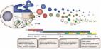
          Extracellular vesicles are nanoparticles produced by cells. They are composed of cellular membrane with associated membrane proteins that surrounds an aqueous core containing soluble molecules such as proteins and nucleic acids, like miRNA and mRNA. They are important in many physiological and pathological processes as they can transfer biological molecules from producer cells to acceptor cells. Preparation of the niche for cancer metastasis, stimulation of tissue regeneration and orchestration of the immune response are examples of the diverse processes in which extracellular vesicles have been implicated. As a result, these vesicles have formed a source of inspiration for many scientific fields. They could be used, for example, as liquid biopsies in diagnostics, as therapeutics in regenerative medicine, or as drug delivery vehicles for transport of medicines. In this Account, we focus on drug delivery applications.

          As we learn more and more about these vesicles, the complexity increases. What originally appeared to be a relatively uniform population of cellular vesicles is increasingly subdivided into different subsets. Cells make various distinct vesicle types whose physicochemical aspects and composition is influenced by parental cell type, cellular activation state, local microenvironment, biogenesis pathway, and intracellular cargo sorting routes. It has proven difficult to assess the effects of changes in production protocol on the characteristics of the cell-derived vesicle population. On top of that, each isolation method for vesicles necessarily enriches certain vesicle classes and subpopulations while depleting others. Also, each method is associated with a varying degree of vesicle purity and concomitant coisolation of nonvesicular material. What emerges is a staggering heterogeneity. This constitutes one of the main challenges of the field as small changes in production and isolation protocols may have large impact on the vesicle characteristics and on subsequent vesicle activity.

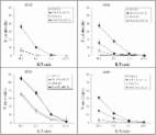
          We try to meet this challenge by careful experimental design and development of tools that enable robust readouts. By engineering the surface and cargo of extracellular vesicles through chemical and biological techniques, favorable characteristics can be enforced while unfavorable qualities can be overruled or masked. This is coupled to the precise evaluation of the interaction of extracellular vesicles with cells to determine the extracellular vesicle uptake routes and intracellular routing. Sensitive reporter assays enable reproducible analysis of functional delivery.

          This systematic evaluation and optimization of extracellular vesicles improves our insight into the critical determinants of extracellular vesicle activity and should improve translation into clinical application of engineered extracellular vesicles as a new class of drug delivery systems.

          Related collections

          Most cited references27

          • Record: found
          • Abstract: found
          • Article: found
          Is Open Access

          In Vivo Imaging Reveals Extracellular Vesicle-Mediated Phenocopying of Metastatic Behavior

          Summary Most cancer cells release heterogeneous populations of extracellular vesicles (EVs) containing proteins, lipids, and nucleic acids. In vitro experiments showed that EV uptake can lead to transfer of functional mRNA and altered cellular behavior. However, similar in vivo experiments remain challenging because cells that take up EVs cannot be discriminated from non-EV-receiving cells. Here, we used the Cre-LoxP system to directly identify tumor cells that take up EVs in vivo. We show that EVs released by malignant tumor cells are taken up by less malignant tumor cells located within the same and within distant tumors and that these EVs carry mRNAs involved in migration and metastasis. By intravital imaging, we show that the less malignant tumor cells that take up EVs display enhanced migratory behavior and metastatic capacity. We postulate that tumor cells locally and systemically share molecules carried by EVs in vivo and that this affects cellular behavior.
            Bookmark
            • Record: found
            • Abstract: found
            • Article: found
            Is Open Access

            Extracellular Vesicle Heterogeneity: Subpopulations, Isolation Techniques, and Diverse Functions in Cancer Progression

            Cells release membrane enclosed nano-sized vesicles termed extracellular vesicles (EVs) that function as mediators of intercellular communication by transferring biological information between cells. Tumor-derived EVs have emerged as important mediators in cancer development and progression, mainly through transfer of their bioactive content which can include oncoproteins, oncogenes, chemokine receptors, as well as soluble factors, transcripts of proteins and miRNAs involved in angiogenesis or inflammation. This transfer has been shown to influence the metastatic behavior of primary tumors. Moreover, tumor-derived EVs have been shown to influence distant cellular niches, establishing favorable microenvironments that support growth of disseminated cancer cells upon their arrival at these pre-metastatic niches. It is generally accepted that cells release a number of major EV populations with distinct biophysical properties and biological functions. Exosomes, microvesicles, and apoptotic bodies are EV populations most widely studied and characterized. They are discriminated based primarily on their intracellular origin. However, increasing evidence suggests that even within these EV populations various subpopulations may exist. This heterogeneity introduces an extra level of complexity in the study of EV biology and function. For example, EV subpopulations could have unique roles in the intricate biological processes underlying cancer biology. Here, we discuss current knowledge regarding the role of subpopulations of EVs in cancer development and progression and highlight the relevance of EV heterogeneity. The position of tetraspanins and integrins therein will be highlighted. Since addressing EV heterogeneity has become essential for the EV field, current and novel techniques for isolating EV subpopulations will also be discussed. Further dissection of EV heterogeneity will advance our understanding of the critical roles of EVs in health and disease.
              Bookmark
              • Record: found
              • Abstract: found
              • Article: found
              Is Open Access

              A phase I study of dexosome immunotherapy in patients with advanced non-small cell lung cancer

              Background There is a continued need to develop more effective cancer immunotherapy strategies. Exosomes, cell-derived lipid vesicles that express high levels of a narrow spectrum of cell proteins represent a novel platform for delivering high levels of antigen in conjunction with costimulatory molecules. We performed this study to test the safety, feasibility and efficacy of autologous dendritic cell (DC)-derived exosomes (DEX) loaded with the MAGE tumor antigens in patients with non-small cell lung cancer (NSCLC). Methods This Phase I study enrolled HLA A2+ patients with pre-treated Stage IIIb (N = 4) and IV (N = 9) NSCLC with tumor expression of MAGE-A3 or A4. Patients underwent leukapheresis to generate DC from which DEX were produced and loaded with MAGE-A3, -A4, -A10, and MAGE-3DPO4 peptides. Patients received 4 doses of DEX at weekly intervals. Results Thirteen patients were enrolled and 9 completed therapy. Three formulations of DEX were evaluated; all were well tolerated with only grade 1–2 adverse events related to the use of DEX (injection site reactions (N = 8), flu like illness (N = 1), and peripheral arm pain (N = 1)). The time from the first dose of DEX until disease progression was 30 to 429+ days. Three patients had disease progression before the first DEX dose. Survival of patients after the first DEX dose was 52–665+ days. DTH reactivity against MAGE peptides was detected in 3/9 patients. Immune responses were detected in patients as follows: MAGE-specific T cell responses in 1/3, increased NK lytic activity in 2/4. Conclusion Production of the DEX vaccine was feasible and DEX therapy was well tolerated in patients with advanced NSCLC. Some patients experienced long term stability of disease and activation of immune effectors
                Bookmark

                Author and article information

                Journal
                Acc Chem Res
                Acc. Chem. Res
                ar
                achre4
                Accounts of Chemical Research
                American Chemical Society
                0001-4842
                1520-4898
                05 June 2019
                16 July 2019
                : 52
                : 7
                : 1761-1770
                Affiliations
                []Laboratory Clinical Chemistry & Hematology, University Medical Center Utrecht, Utrecht University , Heidelberglaan 100, 3584 CX Utrecht, The Netherlands
                []Experimental Cardiology, University Medical Center Utrecht, Utrecht University , Heidelberglaan 100, 3584 CX Utrecht, The Netherlands
                [§ ]University Medical Center Utrecht Regenerative Medicine Center, University Medical Center Utrecht, Utrecht University , Heidelberglaan 100, 3584 CX Utrecht, The Netherlands
                Author notes
                [* ]R.S. E-mail: R.Schiffelers@ 123456umcutrecht.nl . Phone: +31 88 7556512.
                Article
                10.1021/acs.accounts.9b00109
                6639984
                31181910
                9d3a292e-dc1f-4178-8dff-6faf54cab203
                Copyright © 2019 American Chemical Society

                This is an open access article published under a Creative Commons Non-Commercial No Derivative Works (CC-BY-NC-ND) Attribution License, which permits copying and redistribution of the article, and creation of adaptations, all for non-commercial purposes.

                History
                : 02 March 2019
                Categories
                Article
                Custom metadata
                ar9b00109
                ar-2019-00109z

                General chemistry
                General chemistry

                Comments

                Comment on this article