Inviting an author to review:
Find an author and click ‘Invite to review selected article’ near their name.
Search for authorsSearch for similar articles
23
views
0
recommends
+1 Recommend
0 collections
    0
    shares
      • Record: found
      • Abstract: found
      • Article: found
      Is Open Access

      Echocardiographic Assessment of Cardiac Function by Conventional and Speckle‐Tracking Echocardiography in Dogs with Patent Ductus Arteriosus

      research-article

      Read this article at

      Bookmark
          There is no author summary for this article yet. Authors can add summaries to their articles on ScienceOpen to make them more accessible to a non-specialist audience.

          Abstract

          Background

          Patent ductus arteriosus ( PDA) is one of the most common congenital heart defects in dogs. Advanced echocardiographic techniques such as speckle‐tracking echocardiography ( STE) have not been extensively used to evaluate cardiac function in affected dogs.

          Hypothesis

          Advanced echocardiographic techniques are more sensitive than standard echocardiographic techniques in analyzing systolic function in dogs with PDA.

          Animals

          Forty‐four client‐owned dogs: 34 dogs with PDA (preoperative evaluation) and 10 healthy sex‐ and weight‐matched controls.

          Methods

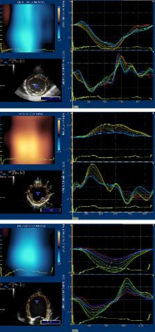
          Prospective study. Dogs were recruited over a 2‐year period. Complete echocardiographic evaluation was performed, including conventional (end‐diastolic volumes indexed to body surface area in B and M‐mode [ EDVI B /M], end‐systolic volumes indexed to body surface area in B and M‐mode [ ESVI B /M], allometric scaling in diastole and systole [AlloD/S], pulmonary flow to systemic flow [Qp/Qs], ejection fraction [ EF] and fractional shortening [ FS]) and speckle‐tracking echocardiography ([ STE]: global longitudinal, radial and circumferential strain [S] and strain rate [ SR]).

          Results

          Dogs with PDA had significantly different EDVI B /M, ESVI B /M, AlloD/S, Qp/Qs and all STE‐derived parameters (global longitudinal S and SR, global circumferential S and SR, global radial S and SR)compared to healthy dogs. No correlation was found between standard techniques ( EDVI B /M, ESVI B /M, AlloD/S, Qp/Qs) and STE‐derived parameters (global longitudinal, circumferential and radial S and SR).

          Conclusion and Clinical Importance

          Conventional parameters routinely used to assess systolic function ( EF and FS) were not different between the groups; STE‐derived parameters identified subtle changes in cardiac systolic function and contractility between the 2 groups of dogs. Based on these findings, STE may be a more appropriate tool to assess cardiac contractility in dogs with PDA.

          Related collections

          Most cited references32

          • Record: found
          • Abstract: found
          • Article: not found

          Definitions for a common standard for 2D speckle tracking echocardiography: consensus document of the EACVI/ASE/Industry Task Force to standardize deformation imaging.

          Recognizing the critical need for standardization in strain imaging, in 2010, the European Association of Echocardiography (now the European Association of Cardiovascular Imaging, EACVI) and the American Society of Echocardiography (ASE) invited technical representatives from all interested vendors to participate in a concerted effort to reduce intervendor variability of strain measurement. As an initial product of the work of the EACVI/ASE/Industry initiative to standardize deformation imaging, we prepared this technical document which is intended to provide definitions, names, abbreviations, formulas, and procedures for calculation of physical quantities derived from speckle tracking echocardiography and thus create a common standard.
            Bookmark
            • Record: found
            • Abstract: found
            • Article: not found

            Recommendations for standards in transthoracic two-dimensional echocardiography in the dog and cat. Echocardiography Committee of the Specialty of Cardiology, American College of Veterinary Internal Medicine.

            Recommendations are presented for standardized imaging planes and display conventions for two-dimensional echocardiography in the dog and cat. Three transducer locations ("windows") provide access to consistent imaging planes: the right parasternal location, the left caudal (apical) parasternal location, and the left cranial parasternal location. Recommendations for image display orientations are very similar to those for comparable human cardiac images, with the heart base or cranial aspect of the heart displayed to the examiner's right on the video display. From the right parasternal location, standard views include a long-axis four-chamber view and a long-axis left ventricular outflow view, and short-axis views at the levels of the left ventricular apex, papillary muscles, chordae tendineae, mitral valve, aortic valve, and pulmonary arteries. From the left caudal (apical) location, standard views include long-axis two-chamber and four-chamber views. From the left cranial parasternal location, standard views include a long-axis view of the left ventricular outflow tract and ascending aorta (with variations to image the right atrium and tricuspid valve, and the pulmonary valve and pulmonary artery), and a short-axis view of the aortic root encircled by the right heart. These images are presented by means of idealized line drawings. Adoption of these standards should facilitate consistent performance, recording, teaching, and communicating results of studies obtained by two-dimensional echocardiography.
              Bookmark
              • Record: found
              • Abstract: found
              • Article: found
              Is Open Access

              Strain and Strain Rate Imaging by Echocardiography – Basic Concepts and Clinical Applicability

              Echocardiographic strain and strain-rate imaging (deformation imaging) is a new non-invasive method for assessment of myocardial function. Due to its ability to differentiate between active and passive movement of myocardial segments, to quantify intraventricular dyssynchrony and to evaluate components of myocardial function, such as longitudinal myocardial shortening, that are not visually assessable, it allows comprehensive assessment of myocardial function and the spectrum of potential clinical applications is very wide. The high sensitivity of both tissue Doppler imaging (TDI) derived and two dimensional (2D) speckle tracking derived myocardial deformation (strain and strain rate) data for the early detection of myocardial dysfunction recommend these new non-invasive diagnostic methods for extensive clinical use. In addition to early detection and quantification of myocardial dysfunction of different etiologies, assessment of myocardial viability, detection of acute allograft rejection and early detection of allograft vasculopathy after heart transplantation, strain and strain rate data are helpful for therapeutic decisions and also useful for follow-up evaluations of therapeutic results in cardiology and cardiac surgery. Strain and strain rate data also provide valuable prognostic information, especially prediction of future reverse remodelling after left ventricular restoration surgery or after cardiac resynchronization therapy and prediction of short and median-term outcome without transplantation or ventricular assist device implantation of patients referred for heart transplantation. The Review explains the fundamental concepts of deformation imaging, describes in a comparative manner the two major deformation imaging methods (TDI-derived and speckle tracking 2D-strain derived) and discusses the clinical applicability of these new echocardiographic tools, which recently have become a subject of great interest for clinicians.
                Bookmark

                Author and article information

                Journal
                J Vet Intern Med
                J. Vet. Intern. Med
                10.1111/(ISSN)1939-1676
                JVIM
                Journal of Veterinary Internal Medicine
                John Wiley and Sons Inc. (Hoboken )
                0891-6640
                1939-1676
                14 May 2016
                May-Jun 2016
                : 30
                : 3 ( doiID: 10.1111/jvim.2016.30.issue-3 )
                : 706-713
                Affiliations
                [ 1 ]Università degli Studi di Milano GallarateItaly
                [ 2 ]Clinica Veterinaria Gran Sasso MilanoItaly
                Author notes
                [*] [* ]Corresponding author: I. Spalla, Università degli Studi di Milano, via padre lega 53, Gallarate, Italy; e‐mail: illispa@ 123456hotmail.com
                Author information
                http://orcid.org/0000-0003-0039-1438
                Article
                JVIM13938
                10.1111/jvim.13938
                4913585
                27177624
                9db62d83-9035-46c7-9fd6-e7a2e484f363
                Copyright © 2016 The Authors. Journal of Veterinary Internal Medicine published by Wiley Periodicals, Inc. on behalf of the American College of Veterinary Internal Medicine.

                This is an open access article under the terms of the Creative Commons Attribution‐NonCommercial License, which permits use, distribution and reproduction in any medium, provided the original work is properly cited and is not used for commercial purposes.

                History
                : 02 July 2015
                : 01 February 2016
                : 02 March 2016
                Page count
                Pages: 8
                Categories
                Standard Article
                SMALL ANIMAL
                Standard Articles
                Cardiology
                Custom metadata
                2.0
                jvim13938
                May/June 2016
                Converter:WILEY_ML3GV2_TO_NLMPMC version:4.9.1 mode:remove_FC converted:17.06.2016

                Veterinary medicine
                congenital heart disease,dogs,strain,strain rate
                Veterinary medicine
                congenital heart disease, dogs, strain, strain rate

                Comments

                Comment on this article