21
views
0
recommends
+1 Recommend
0 collections
    0
    shares
      • Record: found
      • Abstract: found
      • Article: found
      Is Open Access

      Regional Brain Volumes Moderate, but Do Not Mediate, the Effects of Group-Based Exercise Training on Reductions in Loneliness in Older Adults

      research-article

      Read this article at

      Bookmark
          There is no author summary for this article yet. Authors can add summaries to their articles on ScienceOpen to make them more accessible to a non-specialist audience.

          Abstract

          Introduction: Despite the prevalence of and negative health consequences associated with perceived loneliness in older adults, few studies have examined interactions among behavioral, psychosocial, and neural mechanisms. Research suggests that physical activity and improvements in perceived social support and stress are related to reductions in loneliness. Yet, the influence of brain structure on these changes is unknown. The present study examined whether change in regional brain volume mediated the effects of changes in social support and stress on change in perceived loneliness after an exercise intervention. We also examined the extent to which baseline brain volumes moderated the relationship between changes in social support, stress, and loneliness.

          Methods: Participants were 247 older adults (65.4 ± 4.6 years-old) enrolled in a 6-month randomized controlled trial comprised of four exercise conditions: Dance ( n = 69), Strength/Stretching/Stability ( n = 70), Walk ( n = 54), and Walk Plus ( n = 54). All groups met for 1 h, three times weekly. Participants completed questionnaires assessing perceived social support, stress, and loneliness at baseline and post-intervention. Regional brain volumes (amygdala, prefrontal cortex [PFC], hippocampus) before and after intervention were measured with automatic segmentation of each participant's T1-weighted structural MRI. Data were analyzed in a latent modeling framework.

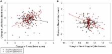
          Results: Perceived social support increased ( p = 0.003), while stress ( p < 0.001), and loneliness ( p = 0.001) decreased over the intervention. Increased social support directly (−0.63, p < 0.01) and indirectly, through decreased stress (−0.10, p = 0.02), predicted decreased loneliness. Changes in amygdala, PFC, and hippocampus volumes were unrelated to change in psychosocial variables (all p ≥ 0.44). However, individuals with larger baseline amygdalae experienced greater decreases in loneliness due to greater reductions in stress (0.35, p = 0.02). Further, individuals with larger baseline PFC volumes experienced greater reductions in stress due to greater increases in social support (−0.47, p = 0.02). No group differences in these pathways were observed.

          Conclusions: The social support environment and resulting reductions in stress, as opposed to exercise mode, may represent important features of exercise programs for improving older adults' perceived loneliness. As amygdala volume has been linked to anxiety, depression and impaired cognitive control processes in the PFC, moderation findings suggest further investigation in this area is warranted.

          Trial Registration: ClinicalTrials.gov identifier NCT01472744 ( https://clinicaltrials.gov/ ct2/show/NCT01472744?term=NCT01472744&rank=1).

          Related collections

          Most cited references35

          • Record: found
          • Abstract: found
          • Article: not found

          Emotional processing in anterior cingulate and medial prefrontal cortex.

          Negative emotional stimuli activate a broad network of brain regions, including the medial prefrontal (mPFC) and anterior cingulate (ACC) cortices. An early influential view dichotomized these regions into dorsal-caudal cognitive and ventral-rostral affective subdivisions. In this review, we examine a wealth of recent research on negative emotions in animals and humans, using the example of fear or anxiety, and conclude that, contrary to the traditional dichotomy, both subdivisions make key contributions to emotional processing. Specifically, dorsal-caudal regions of the ACC and mPFC are involved in appraisal and expression of negative emotion, whereas ventral-rostral portions of the ACC and mPFC have a regulatory role with respect to limbic regions involved in generating emotional responses. Moreover, this new framework is broadly consistent with emerging data on other negative and positive emotions. Published by Elsevier Ltd.
            Bookmark
            • Record: found
            • Abstract: found
            • Article: not found

            Improved Localizadon of Cortical Activity by Combining EEG and MEG with MRI Cortical Surface Reconstruction: A Linear Approach.

            Abstract We describe a comprehensive linear approach to the problem of imaging brain activity with high temporal as well as spatial resolution based on combining EEG and MEG data with anatomical constraints derived from MRI images. The "inverse problem" of estimating the distribution of dipole strengths over the cortical surface is highly underdetermined, even given closely spaced EEG and MEG recordings. We have obtained much better solutions to this problem by explicitly incorporating both local cortical orientation as well as spatial covariance of sources and sensors into our formulation. An explicit polygonal model of the cortical manifold is first constructed as follows: (1) slice data in three orthogonal planes of section (needle-shaped voxels) are combined with a linear deblurring technique to make a single high-resolution 3-D image (cubic voxels), (2) the image is recursively flood-filled to determine the topology of the gray-white matter border, and (3) the resulting continuous surface is refined by relaxing it against the original 3-D gray-scale image using a deformable template method, which is also used to computationally flatten the cortex for easier viewing. The explicit solution to an error minimization formulation of an optimal inverse linear operator (for a particular cortical manifold, sensor placement, noise and prior source covariance) gives rise to a compact expression that is practically computable for hundreds of sensors and thousands of sources. The inverse solution can then be weighted for a particular (averaged) event using the sensor covariance for that event. Model studies suggest that we may be able to localize multiple cortical sources with spatial resolution as good as PET with this technique, while retaining a much finer grained picture of activity over time.
              Bookmark
              • Record: found
              • Abstract: found
              • Article: not found

              Differential aging of the brain: patterns, cognitive correlates and modifiers.

              Deciphering the secret of successful aging depends on understanding the patterns and biological underpinnings of cognitive and behavioral changes throughout adulthood. That task is inseparable from comprehending the workings of the brain, the physical substrate of behavior. In this review, we summarize the extant literature on age-related differences and changes in brain structure, including postmortem and noninvasive magnetic resonance imaging (MRI) studies. Among the latter, we survey the evidence from volumetry, diffusion-tensor imaging, and evaluations of white matter hyperintensities (WMH). Further, we review the attempts to elucidate the mechanisms of age-related structural changes by measuring metabolic markers of aging through magnetic resonance spectroscopy (MRS). We discuss the putative links between the pattern of brain aging and the pattern of cognitive decline and stability. We then present examples of activities and conditions (hypertension, hormone deficiency, aerobic fitness) that may influence the course of normal aging in a positive or negative fashion. Lastly, we speculate on several proposed mechanisms of differential brain aging, including neurotransmitter systems, stress and corticosteroids, microvascular changes, calcium homeostasis, and demyelination.
                Bookmark

                Author and article information

                Contributors
                Journal
                Front Aging Neurosci
                Front Aging Neurosci
                Front. Aging Neurosci.
                Frontiers in Aging Neuroscience
                Frontiers Media S.A.
                1663-4365
                25 April 2017
                2017
                : 9
                : 110
                Affiliations
                [1] 1Department of Kinesiology and Community Health, University of Illinois at Urbana-Champaign Urbana, IL, USA
                [2] 2Beckman Institute, University of Illinois at Urbana-Champaign Urbana, IL, USA
                [3] 3Department of Human Development and Family Studies/Molecular, Cellular and Integrative Neurosciences, Colorado State University Fort Collins, CO, USA
                [4] 4Department of Health and Exercise Science, Wake Forest University Winston-Salem, NC, USA
                [5] 5Office of the Provost, Northeastern University Boston, MA, USA
                Author notes

                Edited by: Philip P. Foster, University of Texas Health Science Center at Houston, USA

                Reviewed by: Jinchong Xu, Johns Hopkins School of Medicine, USA; Laura Lorenzo-López, University of A Coruña, Spain

                *Correspondence: Diane K. Ehlers dkehlers@ 123456illinois.edu
                Article
                10.3389/fnagi.2017.00110
                5403947
                28487648
                a0765ea2-9d7e-42a3-a376-b1ccc94e6c2d
                Copyright © 2017 Ehlers, Daugherty, Burzynska, Fanning, Awick, Chaddock-Heyman, Kramer and McAuley.

                This is an open-access article distributed under the terms of the Creative Commons Attribution License (CC BY). The use, distribution or reproduction in other forums is permitted, provided the original author(s) or licensor are credited and that the original publication in this journal is cited, in accordance with accepted academic practice. No use, distribution or reproduction is permitted which does not comply with these terms.

                History
                : 14 October 2016
                : 04 April 2017
                Page count
                Figures: 4, Tables: 3, Equations: 0, References: 64, Pages: 12, Words: 9564
                Funding
                Funded by: National Institute on Aging 10.13039/100000049
                Award ID: R37 AG025667
                Categories
                Neuroscience
                Original Research

                Neurosciences
                exercise,loneliness,aging,prefrontal cortex,amygdala,social support,stress,psychological
                Neurosciences
                exercise, loneliness, aging, prefrontal cortex, amygdala, social support, stress, psychological

                Comments

                Comment on this article