19
views
0
recommends
+1 Recommend
0 collections
    0
    shares
      • Record: found
      • Abstract: found
      • Article: found
      Is Open Access

      Purpurin Suppresses Candida albicans Biofilm Formation and Hyphal Development

      research-article
      1 , * , 2 , 3
      PLoS ONE
      Public Library of Science

      Read this article at

      Bookmark
          There is no author summary for this article yet. Authors can add summaries to their articles on ScienceOpen to make them more accessible to a non-specialist audience.

          Abstract

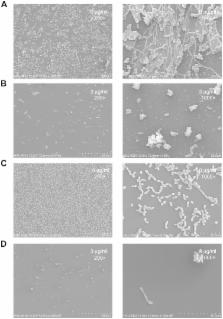
          A striking and clinically relevant virulence trait of the human fungal pathogen Candida albicans is its ability to grow and switch reversibly among different morphological forms. Inhibition of yeast-to-hypha transition in C. albicans represents a new paradigm for antifungal intervention. We have previously demonstrated the novel antifungal activity of purpurin against Candida fungi. In this study, we extended our investigation by examining the in vitro effect of purpurin on C. albicans morphogenesis and biofilms. The susceptibility of C. albicans biofilms to purpurin was examined quantitatively by 2,3-bis(2-methoxy-4-nitro-5-sulfo-phenyl)-2H-tetrazolium-5-carboxanilide reduction assay. Hyphal formation and biofilm ultrastructure were examined qualitatively by scanning electron microscopy (SEM). Quantitative reverse transcription-PCR (qRT-PCR) was used to evaluate the expression of hypha-specific genes and hyphal regulator in purpurin-treated fungal cells. The results showed that, at sub-lethal concentration (3 µg/ml), purpurin blocked the yeast-to-hypha transition under hypha-inducing conditions. Purpurin also inhibited C. albicans biofilm formation and reduced the metabolic activity of mature biofilms in a concentration-dependent manner. SEM images showed that purpurin-treated C. albicans biofilms were scanty and exclusively consisted of aggregates of blastospores. qRT-PCR analyses indicated that purpurin downregulated the expression of hypha-specific genes ( ALS3, ECE1, HWP1, HYR1) and the hyphal regulator RAS1. The data strongly suggested that purpurin suppressed C. albicans morphogenesis and caused distorted biofilm formation. By virtue of the ability to block these two virulence traits in C. albicans, purpurin may represent a potential candidate that deserves further investigations in the development of antifungal strategies against this notorious human fungal pathogen in vivo.

          Related collections

          Most cited references35

          • Record: found
          • Abstract: found
          • Article: not found

          Nonfilamentous C. albicans mutants are avirulent.

          Candida albicans and Saccharomyces cerevisiae switch from a yeast to a filamentous form. In Saccharomyces, this switch is controlled by two regulatory proteins, Ste12p and Phd1p. Single-mutant strains, ste12/ste12 or phd1/phd1, are partially defective, whereas the ste12/ste12 phd1/phd1 double mutant is completely defective in filamentous growth and is noninvasive. The equivalent cph1/cph1 efg1/efg1 double mutant in Candida (Cph1p is the Ste12p homolog and Efg1p is the Phd1p homolog) is also defective in filamentous growth, unable to form hyphae or pseudohyphae in response to many stimuli, including serum or macrophages. This Candida cph1/cph1 efg1/efg1 double mutant, locked in the yeast form, is avirulent in a mouse model.
            Bookmark
            • Record: found
            • Abstract: found
            • Article: not found

            Medicinal plants and antimicrobial activity.

            In the present paper, we analyze the past, present and future of medicinal plants, both as potential antimicrobial crude drugs as well as a source for natural compounds that act as new anti-infection agents. In the past few decades, the search for new anti-infection agents has occupied many research groups in the field of ethnopharmacology. When we reviewed the number of articles published on the antimicrobial activity of medicinal plants in PubMed during the period between 1966 and 1994, we found 115; however, in the following decade between 1995 and 2004, this number more than doubled to 307. In the studies themselves one finds a wide range of criteria. Many focus on determining the antimicrobial activity of plant extracts found in folk medicine, essential oils or isolated compounds such as alkaloids, flavonoids, sesquiterpene lactones, diterpenes, triterpenes or naphtoquinones, among others. Some of these compounds were isolated or obtained by bio-guided isolation after previously detecting antimicrobial activity on the part of the plant. A second block of studies focuses on the natural flora of a specific region or country; the third relevant group of papers is made up of specific studies of the activity of a plant or principle against a concrete pathological microorganism. Some general considerations must be established for the study of the antimicrobial activity of plant extracts, essential oils and the compounds isolated from them. Of utmost relevance is the definition of common parameters, such as plant material, techniques employed, growth medium and microorganisms tested.
              Bookmark
              • Record: found
              • Abstract: found
              • Article: not found

              Antifungal agents: mode of action, mechanisms of resistance, and correlation of these mechanisms with bacterial resistance.

              The increased use of antibacterial and antifungal agents in recent years has resulted in the development of resistance to these drugs. The significant clinical implication of resistance has led to heightened interest in the study of antimicrobial resistance from different angles. Areas addressed include mechanisms underlying this resistance, improved methods to detect resistance when it occurs, alternate options for the treatment of infections caused by resistant organisms, and strategies to prevent and control the emergence and spread of resistance. In this review, the mode of action of antifungals and their mechanisms of resistance are discussed. Additionally, an attempt is made to discuss the correlation between fungal and bacterial resistance. Antifungals can be grouped into three classes based on their site of action: azoles, which inhibit the synthesis of ergosterol (the main fungal sterol); polyenes, which interact with fungal membrane sterols physicochemically; and 5-fluorocytosine, which inhibits macromolecular synthesis. Many different types of mechanisms contribute to the development of resistance to antifungals. These mechanisms include alteration in drug target, alteration in sterol biosynthesis, reduction in the intercellular concentration of target enzyme, and overexpression of the antifungal drug target. Although the comparison between the mechanisms of resistance to antifungals and antibacterials is necessarily limited by several factors defined in the review, a correlation between the two exists. For example, modification of enzymes which serve as targets for antimicrobial action and the involvement of membrane pumps in the extrusion of drugs are well characterized in both the eukaryotic and prokaryotic cells.
                Bookmark

                Author and article information

                Contributors
                Role: Editor
                Journal
                PLoS One
                PLoS ONE
                plos
                plosone
                PLoS ONE
                Public Library of Science (San Francisco, USA )
                1932-6203
                2012
                30 November 2012
                : 7
                : 11
                : e50866
                Affiliations
                [1 ]Oral BioSciences, Faculty of Dentistry, The University of Hong Kong, Hong Kong, China
                [2 ]Division of Pharmaceutics, College of Pharmacy, The University of Texas at Austin, Austin, Texas, United States of America
                [3 ]School of Life Sciences, The Chinese University of Hong Kong, Hong Kong, China
                David Geffen School of Medicine at University of California Los Angeles, United States of America
                Author notes

                Competing Interests: The authors have declared that no competing interests exist.

                Conceived and designed the experiments: PWKT WPF. Performed the experiments: PWKT HMHNB. Analyzed the data: PWKT HMHNB WPF. Contributed reagents/materials/analysis tools: PWKT WPF. Wrote the paper: PWKT WPF.

                Article
                PONE-D-12-11065
                10.1371/journal.pone.0050866
                3511323
                23226409
                a23bcce7-d11f-4302-a4eb-bcc056d54c6e
                Copyright @ 2012

                This is an open-access article distributed under the terms of the Creative Commons Attribution License, which permits unrestricted use, distribution, and reproduction in any medium, provided the original author and source are credited.

                History
                : 17 April 2012
                : 29 October 2012
                Page count
                Pages: 8
                Funding
                This work was partially supported by the Seeding Funding Programme for Basic Research from The University of Hong Kong (Project No. 201003159008 to PWKT), by the Research Fund for the Control of Infectious Diseases, the Food and Health Bureau of the Government of the HKSAR (RFCID 11100992 & 11101122 to PWKT). No additional external funding received for this study.
                Categories
                Research Article
                Biology
                Microbiology
                Mycology
                Fungi
                Yeast
                Pathogenesis
                Model Organisms
                Yeast and Fungal Models
                Candida Albicans
                Molecular Cell Biology
                Gene Expression
                DNA transcription
                Nucleic Acids
                RNA
                RNA synthesis
                Signal Transduction
                Signaling Cascades
                MAPK signaling cascades
                PKA signaling cascade
                Cell Adhesion
                Cell Growth

                Uncategorized
                Uncategorized

                Comments

                Comment on this article