18
views
0
recommends
+1 Recommend
0 collections
    0
    shares
      • Record: found
      • Abstract: found
      • Article: found
      Is Open Access

      Teratocarcinomas Arising from Allogeneic Induced Pluripotent Stem Cell-Derived Cardiac Tissue Constructs Provoked Host Immune Rejection in Mice

      research-article

      Read this article at

      Bookmark
          There is no author summary for this article yet. Authors can add summaries to their articles on ScienceOpen to make them more accessible to a non-specialist audience.

          Abstract

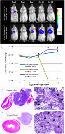
          Transplantation of induced pluripotent stem cell-derived cardiac tissue constructs is a promising regenerative treatment for cardiac failure: however, its tumourigenic potential is concerning. We hypothesised that the tumourigenic potential may be eliminated by the host immune response after allogeneic cell transplantation. Scaffold-free iPSC-derived cardaic tissue sheets of C57BL/6 mouse origin were transplanted into the cardiac surface of syngeneic C57BL/6 mice and allogeneic BALB/c mice with or without tacrolimus injection. Syngeneic mice and tacrolimus-injected immunosuppressed allogeneic mice formed teratocarcinomas with identical phenotypes, characteristic, and time courses, as assessed by imaging tools including 18F-fluorodeoxyglucose-positron emission tomography. In contrast, temporarily immunosuppressed allogeneic mice, following cessation of tacrolimus injection displayed diminished progression of the teratocarcinoma, accompanied by an accumulation of CD4/CD8-positive T cells, and finally achieved complete elimination of the teratocarcinoma. Our results indicated that malignant teratocarcinomas arising from induced pluripotent stem cell-derived cardiac tissue constructs provoked T cell-related host immune rejection to arrest tumour growth in murine allogeneic transplantation models.

          Related collections

          Most cited references18

          • Record: found
          • Abstract: not found
          • Article: not found

          Heart failure.

            Bookmark
            • Record: found
            • Abstract: found
            • Article: not found

            Performance evaluation of the inveon dedicated PET preclinical tomograph based on the NEMA NU-4 standards.

            The Inveon dedicated PET (DPET) scanner is the latest generation of preclinical PET systems devoted to high-resolution and high-sensitivity murine model imaging. In this study, we report on its performance based on the National Electrical Manufacturers Association (NEMA) NU-4 standards. The Inveon DPET consists of 64 lutetium oxyorthosilicate block detectors arranged in 4 contiguous rings, with a 16.1-cm ring diameter and a 12.7-cm axial length. Each detector block consists of a 20 x 20 lutetium oxyorthosilicate crystal array of 1.51 x 1.51 x 10.0 mm elements. The scintillation light is transmitted to position-sensitive photomultiplier tubes via optical light guides. Energy resolution, spatial resolution, sensitivity, scatter fraction, and counting-rate performance were evaluated. The NEMA NU-4 image-quality phantom and a healthy mouse injected with (18)F-FDG and (18)F(-) were scanned to evaluate the imaging capability of the Inveon DPET. The energy resolution at 511 keV was 14.6% on average for the entire system. In-plane radial and tangential resolutions reconstructed with Fourier rebinning and filtered backprojection algorithms were below 1.8-mm full width at half maximum (FWHM) at the center of the field of view. The radial and tangential resolution remained under 2.0 mm, and the axial resolution remained under 2.5-mm FWHM within the central 4-cm diameter of the field of view. The absolute sensitivity of the system was 9.3% for an energy window of 250-625 keV and a timing window of 3.432 ns. At a 350- to 625-keV energy window and a 3.432-ns timing window, the peak noise equivalent counting rate was 1,670 kcps at 130 MBq for the mouse-sized phantom and 590 kcps at 110 MBq for the rat-sized phantom. The scatter fractions at the same acquisition settings were 7.8% and 17.2% for the mouse- and rat-sized phantoms, respectively. The mouse image-quality phantom results demonstrate that for typical mouse acquisitions, the image quality correlates well with the measured performance parameters in terms of image uniformity, recovery coefficients, attenuation, and scatter corrections. The Inveon system, compared with previous generations of preclinical PET systems from the same manufacturer, shows significantly improved energy resolution, sensitivity, axial coverage, and counting-rate capabilities. The performance of the Inveon is suitable for successful murine model imaging experiments.
              Bookmark
              • Record: found
              • Abstract: found
              • Article: not found

              Differentiation of malignant and benign cardiac tumors using 18F-FDG PET/CT.

              In the diagnostic algorithm of cardiac tumors, the noninvasive determination of malignancy and metastatic spread is of major interest to stratify patients and to select and monitor therapies. In the diagnostic work-up, morphologic imaging modalities such as echocardiography or magnetic resonance tomography offer information on, for example, size, invasiveness, and vascularization. However, preoperative assessment of malignancy may be unsatisfactory. The aim of this study was to evaluate the diagnostic value of (18)F-FDG PET and the incremental diagnostic value of an optimized CT score in this clinical scenario. (18)F-FDG PET/CT scans (whole-body imaging with low-dose CT) of 24 consecutive patients with newly diagnosed cardiac tumors were analyzed (11 men, 13 women; mean age ± SD, 59 ± 13 y). The maximum standardized uptake values (SUV(max)) of the tumors were measured. Patients were divided into 2 groups: benign cardiac tumors (n = 7) and malignant cardiac tumors (n = 17) (cardiac primaries [n = 8] and metastases [n = 9]). SUV(max) was compared between the 2 groups. Results were compared with contrast-enhanced CT, using standardized criteria of malignancy. Histology served as ground truth. Mean SUV(max) was 2.8 ± 0.6 in benign cardiac tumors and significantly higher both in malignant primary and in secondary cardiac tumors (8.0 ± 2.1 and 10.8 ± 4.9, P < 0.01). Malignancy was determined with a sensitivity of 100% and specificity of 86% (accuracy, 96%), after a cutoff with high sensitivity (SUV(max) of 3.5) was chosen to avoid false-negatives. Morphologic imaging reached a sensitivity of 82% and a specificity of 86% (accuracy, 83%). Both false-positive and false-negative decisions in morphology could be corrected in all but 1 case using a metabolic threshold with an SUV(max) of 3.5. In addition, extracardiac tumor manifestations were detected in 4 patients by whole-body (18)F-FDG PET/CT. (18)F-FDG PET/CT can aid the noninvasive preoperative determination of malignancy and may be helpful in detecting metastases of malignant cardiac tumors.
                Bookmark

                Author and article information

                Journal
                Sci Rep
                Sci Rep
                Scientific Reports
                Nature Publishing Group
                2045-2322
                14 January 2016
                2016
                : 6
                : 19464
                Affiliations
                [1 ]Department of Cardiovascular Surgery, Osaka University Graduate School of Medicine , Suita, Osaka, Japan
                [2 ]Department of Nuclear Medicine and Tracer Kinetics, Osaka University Graduate School of Medicine , Suita, Osaka, Japan
                [3 ]Immunology Frontier Research Center, Osaka University , Suita, Osaka, Japan
                [4 ]Department of Diagnostic Pathology, Osaka University Graduate School of Medicine , Suita, Osaka, Japan
                Author notes
                Article
                srep19464
                10.1038/srep19464
                4725880
                26763872
                a2af7695-3437-4065-8fd9-f7e8fbea004a
                Copyright © 2016, Macmillan Publishers Limited

                This work is licensed under a Creative Commons Attribution 4.0 International License. The images or other third party material in this article are included in the article’s Creative Commons license, unless indicated otherwise in the credit line; if the material is not included under the Creative Commons license, users will need to obtain permission from the license holder to reproduce the material. To view a copy of this license, visit http://creativecommons.org/licenses/by/4.0/

                History
                : 22 June 2015
                : 09 December 2015
                Categories
                Article

                Uncategorized
                Uncategorized

                Comments

                Comment on this article