12
views
0
recommends
+1 Recommend
0 collections
    0
    shares
      • Record: found
      • Abstract: found
      • Article: found
      Is Open Access

      4‐1BB Delineates Distinct Activation Status of Exhausted Tumor‐Infiltrating CD8 + T Cells in Hepatocellular Carcinoma

      research-article

      Read this article at

      Bookmark
          There is no author summary for this article yet. Authors can add summaries to their articles on ScienceOpen to make them more accessible to a non-specialist audience.

          Abstract

          Background and Aims

          Targeting costimulatory receptors with agonistic antibodies is a promising cancer immunotherapy option. We aimed to investigate costimulatory receptor expression, particularly 4‐1BB (CD137 or tumor necrosis factor receptor superfamily member 9), on tumor‐infiltrating CD8 + T cells (CD8 + tumor‐infiltrating lymphocytes [TILs]) and its association with distinct T‐cell activation features among exhausted CD8 + TILs in hepatocellular carcinoma (HCC).

          Approach and Results

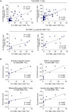
          Tumor tissues, adjacent nontumor tissues, and peripheral blood were collected from HCC patients undergoing surgical resection (n = 79). Lymphocytes were isolated and used for multicolor flow cytometry, RNA‐sequencing, and in vitro functional restoration assays. Among the examined costimulatory receptors, 4‐1BB was most prominently expressed on CD8 + TILs. 4‐1BB expression was almost exclusively detected on CD8 + T cells in the tumor—especially on programmed death 1 (PD‐1) high cells and not PD‐1 int and PD‐1 neg cells. Compared to PD‐1 int and 4‐1BB negPD‐1 high CD8 + TILs, 4‐1BB posPD‐1 high CD8 + TILs exhibited higher levels of tumor reactivity and T‐cell activation markers and significant enrichment for T‐cell activation gene signatures. Per‐patient analysis revealed positive correlations between percentages of 4‐1BB pos cells among CD8 + TILs and levels of parameters of tumor reactivity and T‐cell activation. Among highly exhausted PD‐1 high CD8 + TILs, 4‐1BB pos cells harbored higher proportions of cells with proliferative and reinvigoration potential. Our 4‐1BB–related gene signature predicted survival outcomes of HCC patients in the The Cancer Genome Atlas cohort. 4‐1BB agonistic antibodies enhanced the function of CD8 + TILs and further enhanced the anti‐PD‐1–mediated reinvigoration of CD8 + TILs, especially in cases showing high levels of T‐cell activation.

          Conclusion

          4‐1BB expression on CD8 + TILs represents a distinct activation state among highly exhausted CD8 + T cells in HCC. 4‐1BB costimulation with agonistic antibodies may be a promising strategy for treating HCCs exhibiting prominent T‐cell activation.

          Related collections

          Most cited references19

          • Record: found
          • Abstract: found
          • Article: found
          Is Open Access

          Co-expression of CD39 and CD103 identifies tumor-reactive CD8 T cells in human solid tumors

          Identifying tumor antigen-specific T cells from cancer patients has important implications for immunotherapy diagnostics and therapeutics. Here, we show that CD103+CD39+ tumor-infiltrating CD8 T cells (CD8 TIL) are enriched for tumor-reactive cells both in primary and metastatic tumors. This CD8 TIL subset is found across six different malignancies and displays an exhausted tissue-resident memory phenotype. CD103+CD39+ CD8 TILs have a distinct T-cell receptor (TCR) repertoire, with T-cell clones expanded in the tumor but present at low frequencies in the periphery. CD103+CD39+ CD8 TILs also efficiently kill autologous tumor cells in a MHC-class I-dependent manner. Finally, higher frequencies of CD103+CD39+ CD8 TILs in patients with head and neck cancer are associated with better overall survival. Our data thus describe an approach for detecting tumor-reactive CD8 TILs that will help define mechanisms of existing immunotherapy treatments, and may lead to future adoptive T-cell cancer therapies.
            Bookmark
            • Record: found
            • Abstract: found
            • Article: not found

            The TNF Receptor Superfamily in Co-stimulating and Co-inhibitory Responses.

            Cytokines related to tumor necrosis factor (TNF) provide a communication network essential for coordinating multiple cell types into an effective host defense system against pathogens and malignant cells. The pathways controlled by the TNF superfamily differentiate both innate and adaptive immune cells and modulate stromal cells into microenvironments conducive to host defenses. Members of the TNF receptor superfamily activate diverse cellular functions from the production of type 1 interferons to the modulation of survival of antigen-activated T cells. Here, we focus attention on the subset of TNF superfamily receptors encoded in the immune response locus in chromosomal region 1p36. Recent studies have revealed that these receptors use diverse mechanisms to either co-stimulate or restrict immune responses. Translation of the fundamental mechanisms of TNF superfamily is leading to the design of therapeutics that can alter pathogenic processes in several autoimmune diseases or promote immunity to tumors.
              Bookmark
              • Record: found
              • Abstract: not found
              • Article: not found

              The promise and challenges of immune agonist antibody development in cancer

                Bookmark

                Author and article information

                Contributors
                shwang@amc.seoul.kr
                park3@kaist.ac.kr
                Journal
                Hepatology
                Hepatology
                10.1002/(ISSN)1527-3350
                HEP
                Hepatology (Baltimore, Md.)
                John Wiley and Sons Inc. (Hoboken )
                0270-9139
                1527-3350
                18 October 2019
                March 2020
                : 71
                : 3 ( doiID: 10.1002/hep.v71.3 )
                : 955-971
                Affiliations
                [ 1 ] Graduate School of Medical Science and Engineering Korea Advanced Institute of Science and Technology Daejeon Republic of Korea
                [ 2 ] Biomedical Science and Engineering Interdisciplinary Program Korea Advanced Institute of Science and Technology Daejeon Republic of Korea
                [ 3 ] Department of Pathology, Asan Medical Center University of Ulsan College of Medicine Seoul Republic of Korea
                [ 4 ] Department of Obstetrics and Gynecology Severance Hospital, Yonsei University College of Medicine Seoul Republic of Korea
                [ 5 ] Department of Thoracic and Cardiovascular Surgery Samsung Medical Center, Sungkyunkwan University School of Medicine Seoul Republic of Korea
                [ 6 ] Department of Surgery Severance Hospital, Yonsei University College of Medicine Seoul Republic of Korea
                [ 7 ] Department of Neurosurgery, Severance Hospital Yonsei University College of Medicine Seoul Republic of Korea
                [ 8 ] Department of Surgery, Asan Medical Center University of Ulsan College of Medicine Seoul Republic of Korea
                Author notes
                [*] [* ] Address Correspondence and Reprint Requests to:

                Su‐Hyung Park, Ph.D.

                Laboratory of Translational Immunology and Vaccinology, Graduate School of Medical Science and Engineering, Korea Advanced Institute of Science and Technology

                291 Daehak‐ro

                Daejeon 34141, Republic of Korea

                E‐mail: park3@ 123456kaist.ac.kr

                Tel.: +82‐42‐350‐4248; or

                Shin Hwang, M.D., Ph.D.

                Division of Hepatobiliary Surgery and Liver Transplantation, Department of Surgery, Asan Medical Center, University of Ulsan College of Medicine, Seoul, Republic of Korea

                88, Olympic‐ro 43‐gil

                Songpa‐gu

                Seoul, 05505, Korea

                E‐mail: shwang@ 123456amc.seoul.kr

                Tel.: +82‐2‐3010‐3930

                Author information
                https://orcid.org/0000-0001-6363-7736
                Article
                HEP30881
                10.1002/hep.30881
                7154753
                31353502
                a428edc9-f1bd-495d-9f51-7ce6371c884d
                © 2019 The Authors. Hepatology published by Wiley Periodicals, Inc., on behalf of American Association for the Study of Liver Diseases.

                This is an open access article under the terms of the http://creativecommons.org/licenses/by-nc/4.0/ License, which permits use, distribution and reproduction in any medium, provided the original work is properly cited and is not used for commercial purposes.

                History
                : 12 March 2019
                : 19 July 2019
                Page count
                Figures: 6, Tables: 1, Pages: 17, Words: 18665
                Funding
                Funded by: National Research Foundation of Korea , open-funder-registry 10.13039/501100003725;
                Award ID: NRF‐2016H1A2A1906766
                Funded by: Ministry of Health & Welfare, Republic of Korea
                Award ID: HI15C2859
                Funded by: Deawoong Foundation
                Award ID: DF-201906-0000003
                Categories
                Original Article
                Original Articles
                Hepatobiliary Malignancies
                Custom metadata
                2.0
                March 2020
                Converter:WILEY_ML3GV2_TO_JATSPMC version:5.8.0 mode:remove_FC converted:14.04.2020

                Gastroenterology & Hepatology
                Gastroenterology & Hepatology

                Comments

                Comment on this article