21
views
0
recommends
+1 Recommend
0 collections
    0
    shares
      • Record: found
      • Abstract: found
      • Article: found
      Is Open Access

      A rare anatomic variant of a single-conduit supraclavicular cephalic arch draining into the external jugular vein presenting with recurrent arteriovenous fistula stenosis in a hemodialysis patient

      case-report
      , MBBS , , MBChB, MRCP, , MBBS, MRCP
      Journal of Vascular Surgery Cases and Innovative Techniques
      Elsevier

      Read this article at
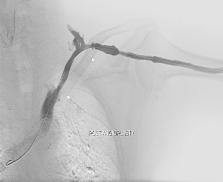
      Bookmark
          There is no author summary for this article yet. Authors can add summaries to their articles on ScienceOpen to make them more accessible to a non-specialist audience.

          Abstract

          The cephalic arch is a common location of stenosis, especially in brachiocephalic fistulas. The cephalic arch has a number of anatomic variations. Cephalic arch stenoses are often resistant and have poor primary patency. Here we describe an unusual case of a hemodialysis patient with a single-conduit supraclavicular cephalic arch draining into the external jugular vein presenting with recurrent cephalic arch stenoses and external jugular vein stenosis. In our view, extrinsic compression by the clavicle may contribute to the high rate of recurrence, the lack of complete dilation of even high-pressure balloons, and a theoretically heightened risk of rupture when cutting balloons are used.

          Related collections

          Most cited references17

          • Record: found
          • Abstract: found
          • Article: not found

          Neointimal hyperplasia in early arteriovenous fistula failure.

          Hemodialysis vascular access dysfunction currently is a huge clinical problem. Although arteriovenous fistulas (AVFs) are the preferred form of permanent dialysis access, they continue to have significant problems with early AVF failure. Although inadequate dilatation of the venous segment was believed to have a role in early AVF failure, the exact pathogenesis of early AVF failure is unknown despite the magnitude of the clinical problem. Case series. Hemodialysis patients. Stenotic venous segments from 4 patients with early AVF failure were subjected to a detailed histological, morphometric, and immunohistochemical analysis. All 4 patients had significant luminal stenosis, primarily as a result of eccentric neointimal hyperplasia. This was confirmed through morphometric analysis, which documented intima-media area and thickness ratios that were greater than unity. Cellular phenotyping studies showed that the majority of cells within the region of neointimal hyperplasia were myofibroblasts, with smaller numbers of contractile smooth muscle cells. We described only a limited number of specimens. We show for the first time that aggressive neointimal hyperplasia is present in venous segment specimens from patients with early AVF failure. Future therapies to address this problem will need to target this pathogenetic pathway.
            Bookmark
            • Record: found
            • Abstract: found
            • Article: not found

            Hemodialysis vascular access morbidity in the United States.

            Extensive morbidity related to hemodialysis vascular access exists among end-stage renal disease (ESRD) patients, but the risk factors for this morbidity have not been extensively studied. Medicare ESRD patient data were obtained from 1984, 1985, and 1986. Hospitalization for vascular access morbidity (ICD-996.1, 996.6, or 996.7) was analyzed among prevalent patients and, using survival analysis, among incident patients to assess sex, age, race, and underlying cause of renal failure as risk factors. We found that 15 to 16% of hospital stays among prevalent ESRD patients were associated with vascular access-related morbidity. Black race, older age, female sex, and diabetes mellitus as a cause of kidney failure were all independent risk factors for access-related morbidity. The rate ratio comparing Blacks to Whites was 1.12 (95% C.I., 1.09, 1.16); > 64 years to 20 to 44 years, 1.53 (1.46, 1.59); men to women, 0.81 (0.79, 0.84); and diabetes to glomerulonephritis, 1.29 (1.24, 1.35). We conclude that hemodialysis vascular access malfunction causes much hospitalization among ESRD patients. Women, Blacks, the elderly, and diabetics appear to be at particularly high risk, and additional studies are needed to understand these patterns.
              Bookmark
              • Record: found
              • Abstract: found
              • Article: not found

              Prevalence and treatment of cephalic arch stenosis in dysfunctional autogenous hemodialysis fistulas.

              Cephalic arch stenosis (CAS) is a recently recognized cause of dysfunction in autogenous hemodialysis fistulas. The prevalence of this lesion among dysfunctional autogenous fistulas is described, as are outcomes after percutaneous therapy.
                Bookmark

                Author and article information

                Contributors
                Journal
                J Vasc Surg Cases Innov Tech
                J Vasc Surg Cases Innov Tech
                Journal of Vascular Surgery Cases and Innovative Techniques
                Elsevier
                2468-4287
                22 March 2017
                March 2017
                22 March 2017
                : 3
                : 1
                : 20-22
                Affiliations
                [1]Khoo Teck Puat Hospital, Singapore
                Author notes
                []Correspondence: Eran Sim Wen Jun, MBBS, 40 Dakota Crescent, 05-13, Singapore 39993940 Dakota Crescent, 05-13399939Singapore Eran_sim@ 123456hotmail.com
                Article
                S2468-4287(16)30102-2
                10.1016/j.jvscit.2016.12.001
                5757772
                a43a4a22-8e2c-4557-bc00-9aed318dc8e0
                © 2016 The Authors

                This is an open access article under the CC BY-NC-ND license (http://creativecommons.org/licenses/by-nc-nd/4.0/).

                History
                : 1 June 2016
                : 13 December 2016
                Categories
                Abdominal aortic aneurysm

                Comments

                Comment on this article