- Record: found
- Abstract: found
- Article: not found
Unusual Location of a Cervical Paraganglioma Between the Thyroid Gland and the Common Carotid Artery: Case Report
letter
Read this article at

There is no author summary for this article yet. Authors can add summaries to their articles on ScienceOpen to make them more accessible to a non-specialist audience.
Abstract
INTRODUCTION
Paragangliomas are rare tumors derived from the extra-adrenal paraganglionic system,
which is composed of cells from the neural crest that are associated with the autonomous
nervous system.1,2 They have been observed at many sites in the head and neck area,
including the carotid body, the jugular-tympanic region, and the vagus nerve.2,3 Unlike
pheochromocytomas, head and neck paragangliomas are usually non-functioning tumors;
in other words, they do not secrete catecholamines, and the primary symptom is a slow
growing mass in the neck.2,4 Paragangliomas arising within the thyroid gland or the
neighboring area are extremely rare, and are believed to derive from the inferior
laryngeal paraganglia.5–9 The laryngeal paraganglia was first described in 1963 by
Watzka,10 who showed the presence of this particular tissue in the upper and anterior
third of ventricular folds. One year later, Kleinsasser11 reported the presence of
paraganglia in the subglottic larynx and named it the inferior laryngeal glomus; this
author also labeled Watzka’s discovery as the superior laryngeal glomus. In anatomical
studies of the human larynges, Zak12 and Lawson13 demonstrated that the position of
the inferior laryngeal paraganglia can vary from patient to patient. They found paraganglionic
tissue to lie between the inferior horn of the thyroid cartilage and the cricoid cartilage,
to lie between the cricoid and the first tracheal ring, or to be in intimate contact
with the medial aspect of the capsule of the thyroid gland. Aberrant positions, such
as in front of the cricoid cartilage and over the cricothyroid membrane, have also
been described.11–13 The association between the inferior laryngeal paraganglia and
the inferior laryngeal nerve and its branches has been well document.11–14
The objective of this paper is to report the diagnosis and treatment of a paraganglioma
located in the anterior visceral cervical space, between the thyroid gland and the
common carotid artery, which during surgery showed no relationship with the recurrent
laryngeal nerve and was easily dissected from the thyroid capsule. The origin of the
reported tumor, which cannot be explained by the previous anatomical descriptions
of the inferior laryngeal paraganglia, is discussed.
CASE DESCRIPTION
A 38-year-old woman complaining of a slowly growing cervical mass was referred to
our clinic. Her personal and family histories were unremarkable. In the physical examination,
a 5-cm, oval-shaped tumor was noted in the position of the right thyroid lobe. The
mass was immobile during deglutition. An ultrasonography suggested a parathyroid mass.
A CT scan was performed and showed a well-defined and highly vascularized tumor located
beneath the right thyroid lobe, which was displaced in the anterior direction (Figure
1). The mass was located between the common carotid artery and the postero-lateral
aspect of the thyroid, and it appeared to share a cleavage plane with the gland. The
patient was submitted to a fine needle aspiration biopsy, and the material was analyzed
by light microscopy. Cytological analysis using Giemsa stain showed numerous cells
with round or oval nuclei, finely granular chromatin, and scanty cytoplasm. The cells
were grouped in single layers around branched capillaries. An immunohistochemical
study was performed on the cell block: it tested positive for chromogranin, synaptophysin,
and vimentin; it tested negative for thyroglobulin, calcitonin, parathyroid hormone,
and cytokeratins. These results suggested a diagnosis of paraganglioma. Urinary tests
for catecholamines were normal.
The patient was then submitted to a cervicotomy through a traditional thyroidectomy
incision. During surgery, a reddish tumor was easily dissected from the postero-lateral
aspect of the right thyroid lobe, which was preserved (Figures 2 and 3). The recurrent
laryngeal nerve was identified and preserved using standard procedures. No relationship
was observed between the tumor and this nerve or any of its branches. No excessive
bleeding or other operative or post-operative complications were observed. The histopathological
examination as well as a new immunohistochemical test with a panel of antibodies confirmed
the diagnosis of paraganglioma (Table 1, Figures 4 and 5). No tumor recurrence or
metastasis was detected six months after surgery, and the patient continues to be
observed in regular follow-up exams.
DISCUSSION
Paragangliomas may theoretically arise at any site where normal paraganglionic tissue
exists. In the head and neck, the carotid body is the most frequently described location
of such tumors, but several other sites have been described in the literature.2,3
Uncommon sites include the laryngeal,14–17 tracheal,18 and thyroid paragangliomas.5–9,19–31
The laryngeal paraganglia has been well described in anatomical studies performed
by German researchers,10,11 and more recently by Zak12 and Lawson.13 The latter studies
reported that the inferior laryngeal paraganglionic tissue may extend to the medial
aspect of the thyroid capsule and may be in contact with the trachea. This finding
explained most cases of thyroid paragangliomas.5–9,19–29,31
The first remarkable observation in the present case was the asymptomatic anterior
neck swelling reported by the patient. This complaint may suggest a benign goiter.
However, the immobility of the mass during deglutition caught our attention as a possible
indicator of a non-thyroidal disease. The imaging tests, particularly the CT scan,
demonstrated clearly that the tumor was not inside the thyroid gland. The fine needle
aspiration biopsy together with an immunohistochemical panel of antibodies was essential
to figure out the diagnosis. Positive results for chromogranin, synaptophysin, and
vimentin, coupled with negative results for thyroglobulin, calcitonin, and cytokeratins
were strongly suggestive of a paraganglioma, as described in the literature.3,8,14,16,22–31
Among the disorders most easily mistaken for cervical paragangliomas, particularly
those arising within the larynx or the thyroid, are the neuroendocrine carcinomas,
such as atypical carcinoids and medullary thyroid carcinoma.14,16,19,22–29,31 As postulated
by Martinez-Madrigal et al.,3 the anti-cytokeratin antibody is the single most useful
marker for differentiating between paraganglioma and neuroendocrine carcinomas. Both
atypical carcinoids and medullary thyroid carcinoma often exhibit diffuse positivity
for cytokeratins. Another disorder that may be confused with paragangliomas is hyalinizing
trabecular adenoma, a benign thyroid neoplasm that shows a prominent nesting pattern
resembling a paraganglioma.24,25,29 This rare entity is usually negative for chromogranin
and positive for thyroglobulin and cytokeratins.
During surgery in the present case, the tumor was easily dissected from the thyroid
gland and the common carotid artery. No relationship was observed between the tumor
and the recurrent laryngeal nerve. Although the patient’s gender (female) and age
(40s and 50s), as well as the laterality of the tumor (right side predominance), favored
the diagnosis of an inferior laryngeal paraganglioma,9,14–16 the site where this tumor
developed could not be explained by previous anatomical descriptions of where the
inferior laryngeal paraganglia was located. The lesion was too far from the carotid
body to be considered a displaced carotid body tumor. To our knowledge, only one prior
report has described a similar localization for a cervical paraganglioma.30 Those
authors described a 3.5-cm paraganglioma located immediately posterior to the left
thyroid lobe, but they could not dissect it from the gland, which was therefore partially
removed. Other previous papers have reported paragangliomas located within the thyroid,5,7–9,21–29,31
between the gland and the trachea,15 and even over the thyroid cartilage.6,20 All
of these locations may be explained by anatomical studies documenting the distribution
of cervical paraganglionic tissue.
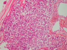
The histopathological and immunohistochemical findings of the specimen confirmed the
diagnosis of paraganglioma in the present case. The classic organoid Zellballen pattern,
composed of nests of chief cells surrounded by sustentacular cells, was observed,
consistent with most previous papers.3,5,7,8,14–19,22–27,29–31 The diagnosis was further
supported by diffuse positivity for chromogranin and synaptophysin in the chief cells,
a focal positivity for S-100 protein in the sustentacular cells, and negativity for
cytokeratins. Despite the absence of histopathological signs of aggressiveness, malignance
cannot be excluded based only on morphological criteria. The diagnosis of a malignant
paraganglioma is based on the detection of regional or distant metastasis and is not
related to the histology of the primary tumor. Hence, all patients submitted to a
paraganglioma resection must be observed in regular follow-up exams to rule out recurrence
or metastasis. However, according to the literature, the ratio of proven cervical
malignant paragangliomas is low: it ranges from 2% to 10% for carotid and vagal body
tumors, to less than 2% for laryngeal paragangliomas.16,19
In conclusion, we have described here an atypical location of a cervical paraganglioma.
This tumor probably originated from an ectopic inferior laryngeal paraganglia or perhaps
from a novel paraganglionic tissue not yet described. This description supports the
idea that the anatomical distribution of human paraganglia is incompletely understood.
We expect that, with the development and spread of immunohistochemical techniques,
new and unusual sites where paragangliomas can arise will be described in the future.
Related collections
Most cited references85
- Record: found
- Abstract: found
- Article: not found
Thyroid paraganglioma: a clinicopathologic and immunohistochemical study of three cases.
J LaGuette, X Matias-Guiu, J Rosai (1997)
- Record: found
- Abstract: found
- Article: not found
Initial work-up and long-term follow-up in patients with phaeochromocytomas and paragangliomas.
- Record: found
- Abstract: found
- Article: not found