49
views
0
recommends
+1 Recommend
0 collections
    0
    shares
      • Record: found
      • Abstract: found
      • Article: found
      Is Open Access

      Perfusion Phantom: An Efficient and Reproducible Method to Simulate Myocardial First-Pass Perfusion Measurements with Cardiovascular Magnetic Resonance

      research-article

      Read this article at

      Bookmark
          There is no author summary for this article yet. Authors can add summaries to their articles on ScienceOpen to make them more accessible to a non-specialist audience.

          Abstract

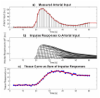
          The aim of this article is to describe a novel hardware perfusion phantom that simulates myocardial first-pass perfusion allowing comparisons between different MR techniques and validation of the results against a true gold standard. MR perfusion images were acquired at different myocardial perfusion rates and variable doses of gadolinium and cardiac output. The system proved to be sensitive to controlled variations of myocardial perfusion rate, contrast agent dose, and cardiac output. It produced distinct signal intensity curves for perfusion rates ranging from 1 to 10 mL/mL/min. Quantification of myocardial blood flow by signal deconvolution techniques provided accurate measurements of perfusion. The phantom also proved to be very reproducible between different sessions and different operators. This novel hardware perfusion phantom system allows reliable, reproducible, and efficient simulation of myocardial first-pass MR perfusion. Direct comparison between the results of image-based quantification and reference values of flow and myocardial perfusion will allow development and validation of accurate quantification methods. Magn Reson Med, 2013. © 2012 Wiley Periodicals, Inc.

          Related collections

          Most cited references31

          • Record: found
          • Abstract: found
          • Article: not found

          Diagnostic performance of stress cardiac magnetic resonance imaging in the detection of coronary artery disease: a meta-analysis.

          The purpose of our study was to conduct an evidence-based evaluation of stress cardiac magnetic resonance imaging (MRI) in the diagnosis of coronary artery disease (CAD). Stress cardiac MRI has recently emerged as a noninvasive method in the detection of CAD, with 2 main techniques in use: 1) perfusion imaging; and 2) stress-induced wall motion abnormalities imaging. We examined studies from January 1990 to January 2007 using MEDLINE and EMBASE. A study was included if it: 1) used stress MRI as a diagnostic test for CAD (> or =50% diameter stenosis); and 2) used catheter X-ray angiography as the reference standard. Thirty-seven studies (2,191 patients) met the inclusion criteria, with 14 datasets (754 patients) using stress-induced wall motion abnormalities imaging and 24 datasets (1,516 patients) using perfusion imaging. Stress-induced wall motion abnormalities imaging demonstrated a sensitivity of 0.83 (95% confidence interval [CI] 0.79 to 0.88) and specificity of 0.86 (95% CI 0.81 to 0.91) on a patient level (disease prevalence = 70.5%). Perfusion imaging demonstrated a sensitivity of 0.91 (95% CI 0.88 to 0.94) and specificity of 0.81 (95% CI 0.77 to 0.85) on a patient level (disease prevalence = 57.4%). In studies with high disease prevalence, stress cardiac MRI, using either technique, demonstrates overall good sensitivity and specificity for the diagnosis of CAD. However, limited data are available regarding use of either technique in populations with low disease prevalence.
            Bookmark
            • Record: found
            • Abstract: found
            • Article: not found

            k-t PCA: temporally constrained k-t BLAST reconstruction using principal component analysis.

            The k-t broad-use linear acquisition speed-up technique (BLAST) has become widespread for reducing image acquisition time in dynamic MRI. In its basic form k-t BLAST speeds up the data acquisition by undersampling k-space over time (referred to as k-t space). The resulting aliasing is resolved in the Fourier reciprocal x-f space (x = spatial position, f = temporal frequency) using an adaptive filter derived from a low-resolution estimate of the signal covariance. However, this filtering process tends to increase the reconstruction error or lower the achievable acceleration factor. This is problematic in applications exhibiting a broad range of temporal frequencies such as free-breathing myocardial perfusion imaging. We show that temporal basis functions calculated by subjecting the training data to principal component analysis (PCA) can be used to constrain the reconstruction such that the temporal resolution is improved. The presented method is called k-t PCA.
              Bookmark
              • Record: found
              • Abstract: found
              • Article: found
              Is Open Access

              Quantification of myocardial perfusion by cardiovascular magnetic resonance

              The potential of contrast-enhanced cardiovascular magnetic resonance (CMR) for a quantitative assessment of myocardial perfusion has been explored for more than a decade now, with encouraging results from comparisons with accepted "gold standards", such as microspheres used in the physiology laboratory. This has generated an increasing interest in the requirements and methodological approaches for the non-invasive quantification of myocardial blood flow by CMR. This review provides a synopsis of the current status of the field, and introduces the reader to the technical aspects of perfusion quantification by CMR. The field has reached a stage, where quantification of myocardial perfusion is no longer a claim exclusive to nuclear imaging techniques. CMR may in fact offer important advantages like the absence of ionizing radiation, high spatial resolution, and an unmatched versatility to combine the interrogation of the perfusion status with a comprehensive tissue characterization. Further progress will depend on successful dissemination of the techniques for perfusion quantification among the CMR community.
                Bookmark

                Author and article information

                Journal
                Magn Reson Med
                Magn Reson Med
                mrm
                Magnetic Resonance in Medicine
                Wiley Subscription Services, Inc., A Wiley Company (Hoboken )
                0740-3194
                1522-2594
                01 March 2013
                24 April 2012
                : 69
                : 3
                : 698-707
                Affiliations
                [1 ]Division of Imaging Sciences, King's College London BHF Centre of Excellence, NIHR Biomedical Research Centre and Wellcome Trust and EPSRC Medical Engineering Centre at Guy's and St. Thomas' NHS Foundation Trust, The Rayne Institute London, United Kingdom
                [2 ]Philips Healthcare, Imaging Systems—MR, Best The Netherlands
                [3 ]Division of Cardiovascular and Neuronal Remodelling, University of Leeds Leeds, United Kingdom
                [4 ]Department of Biomedical Engineering, Biomedical Image Analysis, Eindhoven University of Technology Eindhoven, The Netherlands
                Author notes
                *Correspondence to: Amedeo Chiribiri, M.D., Ph.D., King's College London, Division of Imaging Sciences, The Rayne Institute, 4th Floor Lambeth Wing, St. Thomas' Hospital, London SE1 7EH, United Kingdom. E-mail: amedeo.chiribiri@ 123456kcl.ac.uk
                [†]

                Deceased

                Grant sponsors: Wellcome Trust and EPSRC (The Centre of Excellence in Medical Engineering); Grant number: WT 088641/Z/09/Z; Grant sponsor: British Heart Foundation (BHF); Grant numbers: RE/08/003 and FS/10/029/28253; Grant sponsor: Biomedical Research Centre; Grant numbers: BRC-CTF 196.

                Article
                10.1002/mrm.24299
                3593172
                22532435
                a6429363-22ca-461b-8ec0-c050d3b4e2c4
                Copyright © 2012 Wiley Periodicals, Inc.

                Re-use of this article is permitted in accordance with the Creative Commons Deed, Attribution 2.5, which does not permit commercial exploitation.

                History
                : 03 October 2011
                : 26 March 2012
                : 26 March 2012
                Categories
                Imaging Methodology—Full Paper

                Radiology & Imaging
                first-pass perfusion mr,phantom,gadolinium,3t,heart
                Radiology & Imaging
                first-pass perfusion mr, phantom, gadolinium, 3t, heart

                Comments

                Comment on this article