7
views
0
recommends
+1 Recommend
0 collections
    0
    shares
      • Record: found
      • Abstract: found
      • Article: found
      Is Open Access

      A Novel Method for Classifying Liver and Brain Tumors Using Convolutional Neural Networks, Discrete Wavelet Transform and Long Short-Term Memory Networks

      research-article

      Read this article at

      Bookmark
          There is no author summary for this article yet. Authors can add summaries to their articles on ScienceOpen to make them more accessible to a non-specialist audience.

          Abstract

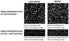
          Rapid classification of tumors that are detected in the medical images is of great importance in the early diagnosis of the disease. In this paper, a new liver and brain tumor classification method is proposed by using the power of convolutional neural network (CNN) in feature extraction, the power of discrete wavelet transform (DWT) in signal processing, and the power of long short-term memory (LSTM) in signal classification. A CNN–DWT–LSTM method is proposed to classify the computed tomography (CT) images of livers with tumors and to classify the magnetic resonance (MR) images of brains with tumors. The proposed method classifies liver tumors images as benign or malignant and then classifies brain tumor images as meningioma, glioma, and pituitary. In the hybrid CNN–DWT–LSTM method, the feature vector of the images is obtained from pre-trained AlexNet CNN architecture. The feature vector is reduced but strengthened by applying the single-level one-dimensional discrete wavelet transform (1-D DWT), and it is classified by training with an LSTM network. Under the scope of the study, images of 56 benign and 56 malignant liver tumors that were obtained from Fırat University Research Hospital were used and a publicly available brain tumor dataset were used. The experimental results show that the proposed method had higher performance than classifiers, such as K-nearest neighbors (KNN) and support vector machine (SVM). By using the CNN–DWT–LSTM hybrid method, an accuracy rate of 99.1% was achieved in the liver tumor classification and accuracy rate of 98.6% was achieved in the brain tumor classification. We used two different datasets to demonstrate the performance of the proposed method. Performance measurements show that the proposed method has a satisfactory accuracy rate at the liver tumor and brain tumor classifying.

          Related collections

          Most cited references53

          • Record: found
          • Abstract: found
          • Article: found
          Is Open Access

          Deep Convolutional Neural Networks for Computer-Aided Detection: CNN Architectures, Dataset Characteristics and Transfer Learning

          Remarkable progress has been made in image recognition, primarily due to the availability of large-scale annotated datasets and deep convolutional neural networks (CNNs). CNNs enable learning data-driven, highly representative, hierarchical image features from sufficient training data. However, obtaining datasets as comprehensively annotated as ImageNet in the medical imaging domain remains a challenge. There are currently three major techniques that successfully employ CNNs to medical image classification: training the CNN from scratch, using off-the-shelf pre-trained CNN features, and conducting unsupervised CNN pre-training with supervised fine-tuning. Another effective method is transfer learning, i.e., fine-tuning CNN models pre-trained from natural image dataset to medical image tasks. In this paper, we exploit three important, but previously understudied factors of employing deep convolutional neural networks to computer-aided detection problems. We first explore and evaluate different CNN architectures. The studied models contain 5 thousand to 160 million parameters, and vary in numbers of layers. We then evaluate the influence of dataset scale and spatial image context on performance. Finally, we examine when and why transfer learning from pre-trained ImageNet (via fine-tuning) can be useful. We study two specific computer-aided detection (CADe) problems, namely thoraco-abdominal lymph node (LN) detection and interstitial lung disease (ILD) classification. We achieve the state-of-the-art performance on the mediastinal LN detection, and report the first five-fold cross-validation classification results on predicting axial CT slices with ILD categories. Our extensive empirical evaluation, CNN model analysis and valuable insights can be extended to the design of high performance CAD systems for other medical imaging tasks.
            Bookmark
            • Record: found
            • Abstract: not found
            • Article: not found

            Decomposition of Hardy Functions into Square Integrable Wavelets of Constant Shape

              Bookmark
              • Record: found
              • Abstract: found
              • Article: found
              Is Open Access

              Enhanced Performance of Brain Tumor Classification via Tumor Region Augmentation and Partition

              Automatic classification of tissue types of region of interest (ROI) plays an important role in computer-aided diagnosis. In the current study, we focus on the classification of three types of brain tumors (i.e., meningioma, glioma, and pituitary tumor) in T1-weighted contrast-enhanced MRI (CE-MRI) images. Spatial pyramid matching (SPM), which splits the image into increasingly fine rectangular subregions and computes histograms of local features from each subregion, exhibits excellent results for natural scene classification. However, this approach is not applicable for brain tumors, because of the great variations in tumor shape and size. In this paper, we propose a method to enhance the classification performance. First, the augmented tumor region via image dilation is used as the ROI instead of the original tumor region because tumor surrounding tissues can also offer important clues for tumor types. Second, the augmented tumor region is split into increasingly fine ring-form subregions. We evaluate the efficacy of the proposed method on a large dataset with three feature extraction methods, namely, intensity histogram, gray level co-occurrence matrix (GLCM), and bag-of-words (BoW) model. Compared with using tumor region as ROI, using augmented tumor region as ROI improves the accuracies to 82.31% from 71.39%, 84.75% from 78.18%, and 88.19% from 83.54% for intensity histogram, GLCM, and BoW model, respectively. In addition to region augmentation, ring-form partition can further improve the accuracies up to 87.54%, 89.72%, and 91.28%. These experimental results demonstrate that the proposed method is feasible and effective for the classification of brain tumors in T1-weighted CE-MRI.
                Bookmark

                Author and article information

                Journal
                Sensors (Basel)
                Sensors (Basel)
                sensors
                Sensors (Basel, Switzerland)
                MDPI
                1424-8220
                28 April 2019
                May 2019
                : 19
                : 9
                : 1992
                Affiliations
                [1 ]Computer Using Department, Besni Vocational School, Adıyaman University, Adıyaman 02300, Turkey
                [2 ]Software Engineering Department, Technology Faculty, Fırat University, Elazığ 23000, Turkey; enginavci@ 123456firat.edu.tr
                Author notes
                [* ]Correspondence: hkutlu@ 123456adiyaman.edu.tr ; Tel.: +90-545-883-0202
                Article
                sensors-19-01992
                10.3390/s19091992
                6540219
                31035406
                a7648cac-705e-4fc8-b3f6-c98cc31c3050
                © 2019 by the authors.

                Licensee MDPI, Basel, Switzerland. This article is an open access article distributed under the terms and conditions of the Creative Commons Attribution (CC BY) license ( http://creativecommons.org/licenses/by/4.0/).

                History
                : 07 March 2019
                : 24 April 2019
                Categories
                Article

                Biomedical engineering
                classification of liver tumor,classification of brain tumor,computer-aided diagnosis,cnn,lstm,dwt,signal classification,feature reduction,biomedical image processing

                Comments

                Comment on this article