5
views
0
recommends
+1 Recommend
0 collections
    0
    shares
      • Record: found
      • Abstract: found
      • Article: found
      Is Open Access

      High-Resolution Free-Breathing Quantitative First-Pass Perfusion Cardiac MR Using Dual-Echo Dixon With Spatio-Temporal Acceleration

      research-article

      Read this article at

      Bookmark
          There is no author summary for this article yet. Authors can add summaries to their articles on ScienceOpen to make them more accessible to a non-specialist audience.

          Abstract

          Introduction

          To develop and test the feasibility of free-breathing (FB), high-resolution quantitative first-pass perfusion cardiac MR (FPP-CMR) using dual-echo Dixon (FOSTERS; Fat-water separation for mOtion-corrected Spatio-TEmporally accelerated myocardial peRfuSion).

          Materials and Methods

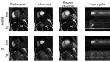
          FOSTERS was performed in FB using a dual-saturation single-bolus acquisition with dual-echo Dixon and a dynamically variable Cartesian k-t undersampling (8-fold) approach, with low-rank and sparsity constrained reconstruction, to achieve high-resolution FPP-CMR images. FOSTERS also included automatic in-plane motion estimation and T 2 * correction to obtain quantitative myocardial blood flow (MBF) maps. High-resolution (1.6 x 1.6 mm 2) FB FOSTERS was evaluated in eleven patients, during rest, against standard-resolution (2.6 x 2.6 mm 2) 2-fold SENSE-accelerated breath-hold (BH) FPP-CMR. In addition, MBF was computed for FOSTERS and spatial wavelet-based compressed sensing (CS) reconstruction. Two cardiologists scored the image quality (IQ) of FOSTERS, CS, and standard BH FPP-CMR images using a 4-point scale (1–4, non-diagnostic – fully diagnostic).

          Results

          FOSTERS produced high-quality images without dark-rim and with reduced motion-related artifacts, using an 8x accelerated FB acquisition. FOSTERS and standard BH FPP-CMR exhibited excellent IQ with an average score of 3.5 ± 0.6 and 3.4 ± 0.6 (no statistical difference, p > 0.05), respectively. CS images exhibited severe artifacts and high levels of noise, resulting in an average IQ score of 2.9 ± 0.5. MBF values obtained with FOSTERS presented a lower variance than those obtained with CS.

          Discussion

          FOSTERS enabled high-resolution FB FPP-CMR with MBF quantification. Combining motion correction with a low-rank and sparsity-constrained reconstruction results in excellent image quality.

          Related collections

          Most cited references55

          • Record: found
          • Abstract: found
          • Article: found

          Magnetic Resonance Perfusion or Fractional Flow Reserve in Coronary Disease

            Bookmark
            • Record: found
            • Abstract: found
            • Article: not found

            Accelerated dynamic MRI exploiting sparsity and low-rank structure: k-t SLR.

            We introduce a novel algorithm to reconstruct dynamic magnetic resonance imaging (MRI) data from under-sampled k-t space data. In contrast to classical model based cine MRI schemes that rely on the sparsity or banded structure in Fourier space, we use the compact representation of the data in the Karhunen Louve transform (KLT) domain to exploit the correlations in the dataset. The use of the data-dependent KL transform makes our approach ideally suited to a range of dynamic imaging problems, even when the motion is not periodic. In comparison to current KLT-based methods that rely on a two-step approach to first estimate the basis functions and then use it for reconstruction, we pose the problem as a spectrally regularized matrix recovery problem. By simultaneously determining the temporal basis functions and its spatial weights from the entire measured data, the proposed scheme is capable of providing high quality reconstructions at a range of accelerations. In addition to using the compact representation in the KLT domain, we also exploit the sparsity of the data to further improve the recovery rate. Validations using numerical phantoms and in vivo cardiac perfusion MRI data demonstrate the significant improvement in performance offered by the proposed scheme over existing methods.
              Bookmark
              • Record: found
              • Abstract: found
              • Article: found
              Is Open Access

              Myocardial perfusion cardiovascular magnetic resonance: optimized dual sequence and reconstruction for quantification

              Background Quantification of myocardial blood flow requires knowledge of the amount of contrast agent in the myocardial tissue and the arterial input function (AIF) driving the delivery of this contrast agent. Accurate quantification is challenged by the lack of linearity between the measured signal and contrast agent concentration. This work characterizes sources of non-linearity and presents a systematic approach to accurate measurements of contrast agent concentration in both blood and myocardium. Methods A dual sequence approach with separate pulse sequences for AIF and myocardial tissue allowed separate optimization of parameters for blood and myocardium. A systems approach to the overall design was taken to achieve linearity between signal and contrast agent concentration. Conversion of signal intensity values to contrast agent concentration was achieved through a combination of surface coil sensitivity correction, Bloch simulation based look-up table correction, and in the case of the AIF measurement, correction of T2* losses. Validation of signal correction was performed in phantoms, and values for peak AIF concentration and myocardial flow are provided for 29 normal subjects for rest and adenosine stress. Results For phantoms, the measured fits were within 5% for both AIF and myocardium. In healthy volunteers the peak [Gd] was 3.5 ± 1.2 for stress and 4.4 ± 1.2 mmol/L for rest. The T2* in the left ventricle blood pool at peak AIF was approximately 10 ms. The peak-to-valley ratio was 5.6 for the raw signal intensities without correction, and was 8.3 for the look-up-table (LUT) corrected AIF which represents approximately 48% correction. Without T2* correction the myocardial blood flow estimates are overestimated by approximately 10%. The signal-to-noise ratio of the myocardial signal at peak enhancement (1.5 T) was 17.7 ± 6.6 at stress and the peak [Gd] was 0.49 ± 0.15 mmol/L. The estimated perfusion flow was 3.9 ± 0.38 and 1.03 ± 0.19 ml/min/g using the BTEX model and 3.4 ± 0.39 and 0.95 ± 0.16 using a Fermi model, for stress and rest, respectively. Conclusions A dual sequence for myocardial perfusion cardiovascular magnetic resonance and AIF measurement has been optimized for quantification of myocardial blood flow. A validation in phantoms was performed to confirm that the signal conversion to gadolinium concentration was linear. The proposed sequence was integrated with a fully automatic in-line solution for pixel-wise mapping of myocardial blood flow and evaluated in adenosine stress and rest studies on N = 29 normal healthy subjects. Reliable perfusion mapping was demonstrated and produced estimates with low variability. Electronic supplementary material The online version of this article (doi:10.1186/s12968-017-0355-5) contains supplementary material, which is available to authorized users.
                Bookmark

                Author and article information

                Contributors
                Journal
                Front Cardiovasc Med
                Front Cardiovasc Med
                Front. Cardiovasc. Med.
                Frontiers in Cardiovascular Medicine
                Frontiers Media S.A.
                2297-055X
                29 April 2022
                2022
                : 9
                : 884221
                Affiliations
                [1] 1Department of Biomedical Engineering, Eindhoven University of Technology , Eindhoven, Netherlands
                [2] 2Department of MR R&D – Clinical Science, Philips Healthcare , Best, Netherlands
                [3] 3Department of Imaging Physics, Magnetic Resonance Systems Lab, Delft University of Technology , Delft, Netherlands
                [4] 4School of Biomedical Engineering and Imaging Sciences, King's College London , London, United Kingdom
                [5] 5Philips Healthcare , Guildford, United Kingdom
                [6] 6Philips Healthcare Iberia , Madrid, Spain
                [7] 7Philips Research , Hamburg, Germany
                [8] 8Division of Cardiovascular Medicine, Department of Medical and Health Sciences, Linkoping University , Linkoping, Sweden
                [9] 9Center for Medical Image Science and Visualization (CMIV), Linkoping University , Linkoping, Sweden
                [10] 10Centre for Marine Sciences (CCMAR) , Faro, Portugal
                Author notes

                Edited by: Sebastian Kelle, Deutsches Herzzentrum Berlin, Germany

                Reviewed by: Vivek Muthurangu, University College London, United Kingdom; Theo Pezel, Hôpital Lariboisière, France; Otávio R. Coelho-Filho, State University of Campinas, Brazil

                *Correspondence: Teresa Correia teresa.correia@ 123456kcl.ac.uk

                This article was submitted to Cardiovascular Imaging, a section of the journal Frontiers in Cardiovascular Medicine

                †These authors have contributed equally to this work

                Article
                10.3389/fcvm.2022.884221
                9099052
                35571164
                a86cb365-162a-41ef-9f11-6379682ce727
                Copyright © 2022 Tourais, Scannell, Schneider, Alskaf, Crawley, Bosio, Sanchez-Gonzalez, Doneva, Schülke, Meineke, Keupp, Smink, Breeuwer, Chiribiri, Henningsson and Correia.

                This is an open-access article distributed under the terms of the Creative Commons Attribution License (CC BY). The use, distribution or reproduction in other forums is permitted, provided the original author(s) and the copyright owner(s) are credited and that the original publication in this journal is cited, in accordance with accepted academic practice. No use, distribution or reproduction is permitted which does not comply with these terms.

                History
                : 25 February 2022
                : 04 April 2022
                Page count
                Figures: 6, Tables: 0, Equations: 1, References: 56, Pages: 10, Words: 7011
                Categories
                Cardiovascular Medicine
                Original Research

                myocardial perfusion,high-resolution,free-breathing,quantitative myocardial blood flow,dixon,motion correction

                Comments

                Comment on this article