2
views
0
recommends
+1 Recommend
0 collections
    0
    shares
      • Record: found
      • Abstract: found
      • Article: found
      Is Open Access

      Noninvasive Imaging Tools in the Diagnosis and Treatment of Skin Cancers

      review-article

      Read this article at

      Bookmark
          There is no author summary for this article yet. Authors can add summaries to their articles on ScienceOpen to make them more accessible to a non-specialist audience.

          Abstract

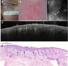
          In the 1980s, the increasing incidence of skin cancers prompted the development of noninvasive medical devices to improve skin cancer diagnosis in daily dermatology practice. As a result of the development of these noninvasive techniques, diagnosis is now established earlier and with better accuracy. These advances are of great benefit to high-risk patients, who previously would have had to undergo several excisions. In this review, we focus on the classic technique of dermoscopy and the more recent digital version, as well as on advanced noninvasive imaging techniques, such as reflectance confocal microscopy and optical coherence tomography. On the basis of their specific features, these noninvasive medical devices can be used not only to diagnose and monitor melanoma and nonmelanoma skin cancers but also to choose the best therapy and follow the patient’s response to treatment in vivo.

          Related collections

          Most cited references44

          • Record: found
          • Abstract: found
          • Article: not found

          Dermoscopy of pigmented skin lesions: results of a consensus meeting via the Internet.

          There is a need for better standardization of the dermoscopic terminology in assessing pigmented skin lesions. The virtual Consensus Net Meeting on Dermoscopy was organized to investigate reproducibility and validity of the various features and diagnostic algorithms. Dermoscopic images of 108 lesions were evaluated via the Internet by 40 experienced dermoscopists using a 2-step diagnostic procedure. The first-step algorithm distinguished melanocytic versus nonmelanocytic lesions. The second step in the diagnostic procedure used 4 algorithms (pattern analysis, ABCD rule, Menzies method, and 7-point checklist) to distinguish melanoma versus benign melanocytic lesions. kappa Values, log odds ratios, sensitivity, specificity, and positive likelihood ratios were estimated for all diagnostic algorithms and dermoscopic features. Interobserver agreement was fair to good for all diagnostic methods, but it was poor for the majority of dermoscopic criteria. Intraobserver agreement was good to excellent for all algorithms and features considered. Pattern analysis allowed the best diagnostic performance (positive likelihood ratio: 5.1), whereas alternative algorithms revealed comparable sensitivity but less specificity. Interobserver agreement on management decisions made by dermoscopy was fairly good (mean kappa value: 0.53). The virtual Consensus Net Meeting on Dermoscopy represents a valid tool for better standardization of the dermoscopic terminology and, moreover, opens up a new territory for diagnosing and managing pigmented skin lesions.
            Bookmark
            • Record: found
            • Abstract: found
            • Article: not found
            Is Open Access

            Meta-analysis of risk factors for cutaneous melanoma: I. Common and atypical naevi.

            A systematic meta-analysis of observational studies of melanoma and one of the most important risk factors, the number of naevi, was conducted in order to clarify aspects of the aetiology of this disease. Following a systematic literature search, relative risks (RRs) were extracted from 46 studies published before September 2002. Dose-response random effects models were used to obtain pooled estimates. Sub-group analysis and meta-regression were carried out to explore sources of between-study variation and bias. Sensitivity analyses investigated the reliability of the results and any publication bias. Number of common naevi was confirmed an important risk factor with a substantially increased risk associated with the presence of 101-120 naevi compared with <15 (pooled Relative Risk (RR) = 6.89; 95% Confidential Interval (CI): 4.63, 10.25) as was the number of atypical naevi (RR = 6.36 95%; CI: 3.80, 10.33; for 5 versus 0). The type of study and source of cases and controls were two study characteristics that significantly influenced the estimates. Case-control studies, in particular when the hospital was the source for cases or controls, appeared to present much lower and more precise estimates than cohort studies.
              Bookmark
              • Record: found
              • Abstract: found
              • Article: not found

              Dermoscopy compared with naked eye examination for the diagnosis of primary melanoma: a meta-analysis of studies performed in a clinical setting.

              Dermoscopy is a noninvasive technique that enables the clinician to perform direct microscopic examination of diagnostic features, not seen by the naked eye, in pigmented skin lesions. Diagnostic accuracy of dermoscopy has previously been assessed in meta-analyses including studies performed in experimental and clinical settings. To assess the diagnostic accuracy of dermoscopy for the diagnosis of melanoma compared with naked eye examination by performing a meta-analysis exclusively on studies performed in a clinical setting. We searched for publications from 1987 to January 2008 and found nine eligible studies. The selected studies compare diagnostic accuracy of dermoscopy with naked eye examination using a valid reference test on consecutive patients with a defined clinical presentation, performed in a clinical setting. Hierarchical summary receiver operator curve analysis was used to estimate the relative diagnostic accuracy for clinical examination with, and without, the use of dermoscopy. We found the relative diagnostic odds ratio for melanoma, for dermoscopy compared with naked eye examination, to be 15.6 [95% confidence interval (CI) 2.9-83.7, P = 0.016]; removal of two outlier studies changed this to 9.0 (95% CI 1.5-54.6, P = 0.03). Dermoscopy is more accurate than naked eye examination for the diagnosis of cutaneous melanoma in suspicious skin lesions when performed in the clinical setting.
                Bookmark

                Author and article information

                Contributors
                +55 513 359 8571 , rbakos@hcpa.edu.br
                +55 119 668 55552 , tatianadermato@gmail.com
                +52 155 304 06609 , roroderm@yahoo.com
                +54 341 523 2323 , gabriel.salerni@e-derma.com.ar
                Journal
                Am J Clin Dermatol
                Am J Clin Dermatol
                American Journal of Clinical Dermatology
                Springer International Publishing (Cham )
                1175-0561
                1179-1888
                30 October 2018
                30 October 2018
                2018
                : 19
                : Suppl 1
                : 3-14
                Affiliations
                [1 ]ISNI 0000 0001 2200 7498, GRID grid.8532.c, Hospital de Clínicas de Porto Alegre, , Universidade Federal do Rio Grande do Sul, ; Rua Ramiro BArcellos 2350, Porto Alegre, RS 90035-007 Brazil
                [2 ]ISNI 0000 0004 0437 1183, GRID grid.413320.7, Skin Cancer Department, , AC Camargo Cancer Center, ; Rua Prof. Antônio Prudente, 211-Liberdade, São Paulo, SP 01509-010 Brazil
                [3 ]ISNI 0000 0001 2159 0001, GRID grid.9486.3, Dermato-Oncology Clinic, Faculty of Medicine, , Universidad Nacional Autónoma de México, ; Circuito Escolar s/n (Edificio Consejo Técnico Facultad de Medicina), Avenida Universidad 3000, Coyoacan, 04510 Mexico City, Mexico
                [4 ]Hospital Provincial del Centenario de Rosario, Argentina, Diagnóstico Médico Oroño, Bv. Oroño 1515, 2000 Rosario, Argentina
                Article
                367
                10.1007/s40257-018-0367-4
                6244601
                30374899
                aa3e135e-95e9-4a7c-8cd3-7f86e2a2981b
                © The Author(s) 2018

                Open AccessThis article is distributed under the terms of the Creative Commons Attribution-NonCommercial 4.0 International License (http://creativecommons.org/licenses/by-nc/4.0/), which permits any noncommercial use, distribution, and reproduction in any medium, provided you give appropriate credit to the original author(s) and the source, provide a link to the Creative Commons license, and indicate if changes were made.

                History
                Categories
                Review Article
                Custom metadata
                © Springer Nature Switzerland AG 2018

                Comments

                Comment on this article