8
views
0
recommends
+1 Recommend
0 collections
    0
    shares
      • Record: found
      • Abstract: found
      • Article: found
      Is Open Access

      Biphasic Alteration of Butyrylcholinesterase (BChE) During Prostate Cancer Development

      research-article

      Read this article at

      Bookmark
          There is no author summary for this article yet. Authors can add summaries to their articles on ScienceOpen to make them more accessible to a non-specialist audience.

          Abstract

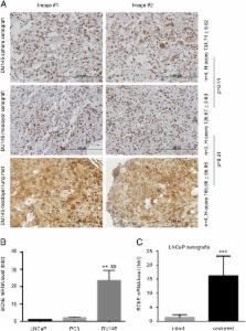
          Butyrylcholinesterase (BChE) is a plasma enzyme that hydrolyzes ghrelin and bioactive esters, suggesting a role in modulating metabolism. Serum BChE is reduced in cancer patients. In prostate cancer (PC), the down-regulation is associated with disease recurrence. Nonetheless, how BChE is expressed in PC and its impact on PC remain unclear. We report here the biphasic changes of BChE expression in PC. In vitro, BChE expression was decreased in more tumorigenic PC stem-like cells (PCSLCs), DU145, and PC3 cells compared to less tumorigenic non-stem PCs and LNCaP cells. On the other hand, BChE was expressed at a higher level in LNCaP cells than immortalized but non-tumorigenic prostate epithelial BPH-1 cells. In vivo, BChE expression was up-regulated in DU145 xenografts compared to LNCaP xenografts; DU145 cell-derived lung metastases displayed comparable levels of BChE as subcutaneous tumors. Furthermore, LNCaP xenografts produced in castrated mice exhibited a significant increase of BChE expression compared to xenografts generated in intact mice. In patients, BChE expression was down-regulated in PCs (n = 340) compared to prostate tissues (n = 86). In two independent PC populations MSKCC (n = 130) and TCGA Provisional (n = 490), BChE mRNA levels were reduced from World Health Organization grade group 1 (WHOGG 1) PCs to WHOGG 3 PCs, followed by a significant increase in WHOGG 5 PCs. The up-regulation was associated with a reduction in disease-free survival ( P = .008). Collectively, we demonstrated for the first time a biphasic alteration of BChE, its down-regulation at early stage of PC and its up-regulation at advanced PC.

          Related collections

          Most cited references43

          • Record: found
          • Abstract: found
          • Article: not found

          PKM2 contributes to cancer metabolism.

          Reprogramming of cell metabolism is essential for tumorigenesis, and is regulated by a complex network, in which PKM2 plays a critical role. PKM2 exists as an inactive monomer, less active dimer and active tetramer. While dimeric PKM2 diverts glucose metabolism towards anabolism through aerobic glycolysis, tetrameric PKM2 promotes the flux of glucose-derived carbons for ATP production via oxidative phosphorylation. Equilibrium of the PKM2 dimers and tetramers is critical for tumorigenesis, and is controlled by multiple factors. The PKM2 dimer also promotes aerobic glycolysis by modulating transcriptional regulation. We will discuss the current understanding of PKM2 in regulating cancer metabolism.
            Bookmark
            • Record: found
            • Abstract: found
            • Article: not found

            Butyrylcholinesterase as a prognostic marker: a review of the literature

            Background Butyrylcholinesterase (BChE) is an α-glycoprotein synthesized in the liver. Its serum level decreases in many clinical conditions such as acute and chronic liver damage, inflammation, injury and infections, and malnutrition. Methods and results This review collects the main evidence on the emerging role of butyrylcholinesterase as a prognostic marker of liver and nonliver diseases as well as a marker of protein-energy malnutrition and obesity. In fact, serum concentrations and BChE activity seem to accurately reflect the availability of amino acidic substrates and/or derangement in protein synthesis due to hepatocellular damage. In cancer, with or without liver impairment, serum BChE levels serve as an accurate functional and prognostic indicator, useful for monitoring clinical and therapeutic interventions according to patients’ prognosis. In the absence of inflammation, BChE could also serve as an index of the effectiveness of nutritional support. Conclusions Serum BChE assessment should be included in routine clinical diagnostic procedures to evaluate patient clinical conditions, in particular in cases of inflammation and/or protein-energy malnutrition.
              Bookmark
              • Record: found
              • Abstract: found
              • Article: not found

              Ghrelin degradation by serum and tissue homogenates: identification of the cleavage sites.

              The endogenous ligand for the GH secretagogue receptor is ghrelin, a peptide recently purified from the stomach. Ghrelin is n-octanoylated on the Ser(3) residue, and this modification is essential for its interaction with the receptor. The degradation of ghrelin by rat and human serum, purified commercial enzymes, and tissues homogenates was analyzed by combining HPLC and mass spectrometry. In serum, ghrelin was desoctanoylated, without proteolysis. The desoctanoylation was significantly reduced by phenylmethylsulfonyl fluoride, a serine proteases and esterases inhibitor. In rat serum, the carboxylesterase inhibitor bis-p-nitrophenyl-phosphate totally inhibited ghrelin desoctanoylation, and a correlation was found between ghrelin desoctanoylation and carboxylesterase activity. Moreover, purified carboxylesterase degraded ghrelin. Thus, carboxylesterase could be responsible for ghrelin desoctanoylation in that species. In human serum, ghrelin desoctanoylation was partially inhibited by eserine salicylate and sodium fluoride, two butyrylcholinesterase inhibitors, but not by bis-p-nitrophenyl-phosphate and EDTA. Purified butyrylcholinesterase was able to degrade ghrelin, and there was a correlation between the butyrylcholinesterase and ghrelin desoctanoylation activities in human sera. This suggested that several esterases, including butyrylcholinesterase, contributed to ghrelin desoctanoylation in human serum. In contact with tissues homogenates, ghrelin was degraded by both desoctanoylation and N-terminal proteolysis. We identified five cleavage sites in ghrelin between residues -Ser(2)-(acyl)Ser(3)- (stomach and liver), -(acyl?)Ser(3)-Phe(4)- (stomach, liver, and kidney), -Phe(4)-Leu(5)- (stomach and kidney), -Leu(5)-Ser(6)- and -Pro(7)-Glu(8)- (kidney). In all cases, the resulting fragments were biologically inactive.
                Bookmark

                Author and article information

                Contributors
                Journal
                Transl Oncol
                Transl Oncol
                Translational Oncology
                Neoplasia Press
                1936-5233
                30 June 2018
                August 2018
                30 June 2018
                : 11
                : 4
                : 1012-1022
                Affiliations
                [* ]Division of Nephrology, Department of Medicine, McMaster University, St. Joseph's Hospital, Hamilton, Ontario, Canada
                []the Research Institute of St Joe's Hamilton, St. Joseph's Hospital, Hamilton, Ontario, Canada
                []the Hamilton Center for Kidney Research, St. Joseph's Hospital, Hamilton, Ontario, Canada
                [§ ]Department of Surgery, McMaster University, Hamilton, Ontario, Canada
                []Department of Nephrology, the First Affiliated Hospital of Nanchang University, Jiangxi, China
                [# ]Department of Gastroenterology, Xiangya Hospital, Central South University, Changsha, Hunan, China
                [** ]School of Medicine, the National University of Ireland, Galway, Ireland
                [†† ]Division of Anatomical Pathology, Department of Pathology and Molecular Medicine, McMaster University, Hamilton, Ontario, Canada
                [‡‡ ]Division of Medical Oncology, Department of Oncology, McMaster University, Hamilton, Ontario, Canada
                Author notes
                [* ]Address all correspondence to: Damu Tang, T3310, St. Joseph's Hospital, 50 Charlton Ave East, Hamilton, Ontario, Canada, L8N 4A6. or Pierre Major, 699 Concession Street, Hamilton, Ontario, Canada, L8V 5C2. majorp@ 123456HHSC.CA damut@ 123456mcmaster.ca
                Article
                S1936-5233(18)30188-8
                10.1016/j.tranon.2018.06.003
                6031255
                29966864
                ad485177-bf49-47a4-bfa4-22ffb9f88f37
                © 2018 The Authors

                This is an open access article under the CC BY-NC-ND license (http://creativecommons.org/licenses/by-nc-nd/4.0/).

                History
                : 2 May 2018
                : 31 May 2018
                : 11 June 2018
                Categories
                Original article

                Comments

                Comment on this article